

Transient blockage of the CD11d/CD18 integrin reduces contusion volume and macrophage infiltration after traumatic brain injury in rats
- PMID: 18374312
- PMCID: PMC2435262
- DOI: 10.1016/j.brainres.2008.02.057
Transient blockage of the CD11d/CD18 integrin reduces contusion volume and macrophage infiltration after traumatic brain injury in rats
Abstract
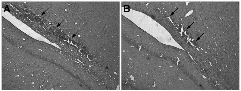
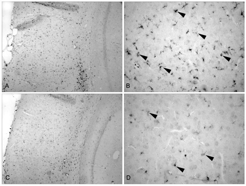
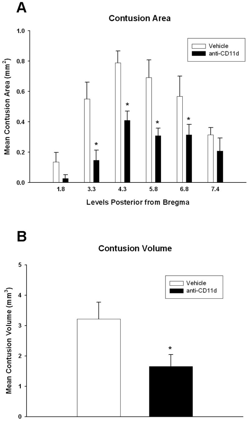
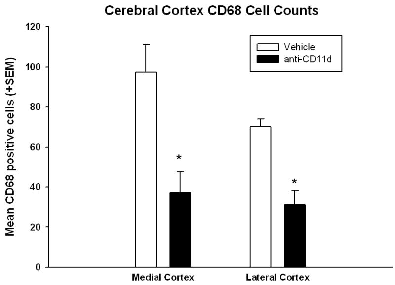
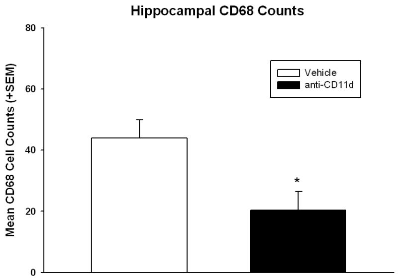
The early inflammatory response to traumatic brain injury (TBI) may result in secondary damage. The purpose of this study was to evaluate the effects of a transient treatment employing a blocking monoclonal antibody (mAb) to the CD11d/CD18 integrin on histopathological outcome and macrophage infiltration following TBI. A parasagittal fluid percussion (FP) brain injury (1.8-2.1 atm) was induced in male Sprague-Dawley rats. Rats were randomized into two trauma groups, treated (N=7) and nontreated (N=8) animals. In the treated group, a mAb to the CD11d subunit of the CD11d/CD18 integrin was administered 30 min, 24 and 48 h after brain injury. Control animals received an isotype-matched irrelevant mAb using the same dose and treatment regimen. At 3 days after TBI, animals were perfusion-fixed for histopathological and immunocytochemical analysis. The anti-CD11d mAb treatment reduced contusion areas as well as overall contusion volume compared to vehicle treated animals. For example, overall contusion volume was reduced from 2.7+/-0.5 mm(3) (mean+/-SEM) to 1.4+/-0.4 with treatment (p<0.05). Immunocytochemical studies identifying CD68 immunoreactive macrophages showed that treatment caused significant attenuation of leukocyte infiltration into the contused cortical areas. These data emphasize the beneficial effects of blocking inflammatory cell recruitment into the injured brain on histopathological outcome following traumatic brain injury.
Figures
Similar articles
-
A CD11d monoclonal antibody treatment reduces tissue injury and improves neurological outcome after fluid percussion brain injury in rats.J Neurotrauma. 2012 Sep 20;29(14):2375-92. doi: 10.1089/neu.2012.2408. Epub 2012 Jul 12. J Neurotrauma. 2012. PMID: 22676851 Free PMC article.
-
CD11d integrin blockade reduces the systemic inflammatory response syndrome after traumatic brain injury in rats.Exp Neurol. 2015 Sep;271:409-22. doi: 10.1016/j.expneurol.2015.07.003. Epub 2015 Jul 11. Exp Neurol. 2015. PMID: 26169930 Free PMC article.
-
The effectiveness of the anti-CD11d treatment is reduced in rat models of spinal cord injury that produce significant levels of intraspinal hemorrhage.Exp Neurol. 2017 Sep;295:125-134. doi: 10.1016/j.expneurol.2017.06.002. Epub 2017 Jun 3. Exp Neurol. 2017. PMID: 28587875
-
Treatment with an anti-CD11d integrin antibody reduces neuroinflammation and improves outcome in a rat model of repeated concussion.J Neuroinflammation. 2013 Feb 15;10:26. doi: 10.1186/1742-2094-10-26. J Neuroinflammation. 2013. PMID: 23414334 Free PMC article.
-
β2 Integrin CD11d/CD18: From Expression to an Emerging Role in Staged Leukocyte Migration.Front Immunol. 2021 Nov 8;12:775447. doi: 10.3389/fimmu.2021.775447. eCollection 2021. Front Immunol. 2021. PMID: 34858434 Free PMC article. Review.
Cited by
-
The Involvement of Pial Microvessels in Leukocyte Invasion after Mild Traumatic Brain Injury.PLoS One. 2016 Dec 28;11(12):e0167677. doi: 10.1371/journal.pone.0167677. eCollection 2016. PLoS One. 2016. PMID: 28030563 Free PMC article.
-
Neuro-glial and systemic mechanisms of pathological responses in rat models of primary blast overpressure compared to "composite" blast.Front Neurol. 2012 Feb 9;3:15. doi: 10.3389/fneur.2012.00015. eCollection 2012. Front Neurol. 2012. PMID: 22403567 Free PMC article.
-
Erythropoietin improves motor and cognitive deficit, axonal pathology, and neuroinflammation in a combined model of diffuse traumatic brain injury and hypoxia, in association with upregulation of the erythropoietin receptor.J Neuroinflammation. 2013 Dec 18;10:156. doi: 10.1186/1742-2094-10-156. J Neuroinflammation. 2013. PMID: 24344874 Free PMC article.
-
Methylene blue attenuates traumatic brain injury-associated neuroinflammation and acute depressive-like behavior in mice.J Neurotrauma. 2015 Jan 15;32(2):127-38. doi: 10.1089/neu.2014.3514. Epub 2014 Nov 13. J Neurotrauma. 2015. PMID: 25070744 Free PMC article.
-
In vivo temporal and spatial profile of leukocyte adhesion and migration after experimental traumatic brain injury in mice.J Neuroinflammation. 2013 Feb 28;10:32. doi: 10.1186/1742-2094-10-32. J Neuroinflammation. 2013. PMID: 23448240 Free PMC article.
References
-
- Barone FC, Feuerstein GZ. Inflammatory mediators and stroke: new opportunities for novel therapeutics. J Cereb Blood Flow Metab. 1999;19:819–34. - PubMed
-
- Becker KJ. Anti-leukocyte antibodies: LeukArrest (Hu23F2G) and Enlimomab (R6.5) in acute stroke. Curr Med Res Opin. 2002;18(Suppl 2):s18–s22. - PubMed
-
- Bethea JR, Dietrich WD. Targeting the host inflammatory response in traumatic spinal cord injury. Curr Opin Neurol. 2002;15:355–360. - PubMed
-
- Bevilacqua MP. Endothelial-leukocyte adhesion molecules. Annu Rev Immunol. 1993;11:767–804. - PubMed
-
- Bye N, Habgood MD, Callaway JK, Malakooti N, Potter A, Kossmann T, Morganti-Kossmann MC. Transient neuroprotection by minocycline following traumatic brain injury is associated with attenuated microglial activation but no changes in cell apoptosis or neutrophil infiltration. Exp Neurol. 2007;204:220–233. - PubMed
Publication types
MeSH terms
Substances
Grants and funding
LinkOut - more resources
Full Text Sources
Other Literature Sources
Research Materials
Miscellaneous
