. 2008 Jun 5;375(2):412-23.
doi: 10.1016/j.virol.2008.02.024. Epub 2008 Apr 18.

Formation of the factory matrix is an important, though not a sufficient function of nonstructural protein mu NS during reovirus infection

Affiliations

Formation of the factory matrix is an important, though not a sufficient function of nonstructural protein mu NS during reovirus infection

Michelle M Arnold et al. Virology. .

Abstract

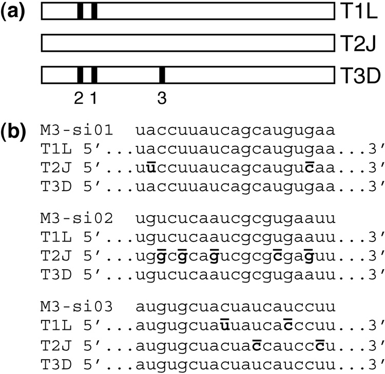
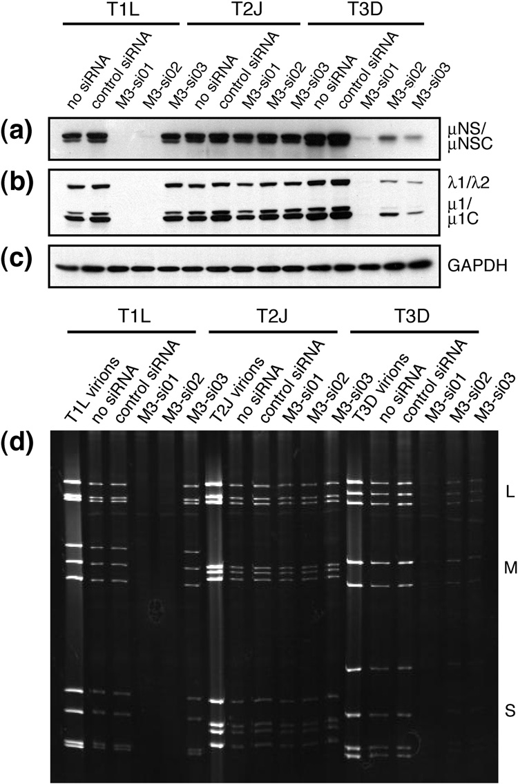
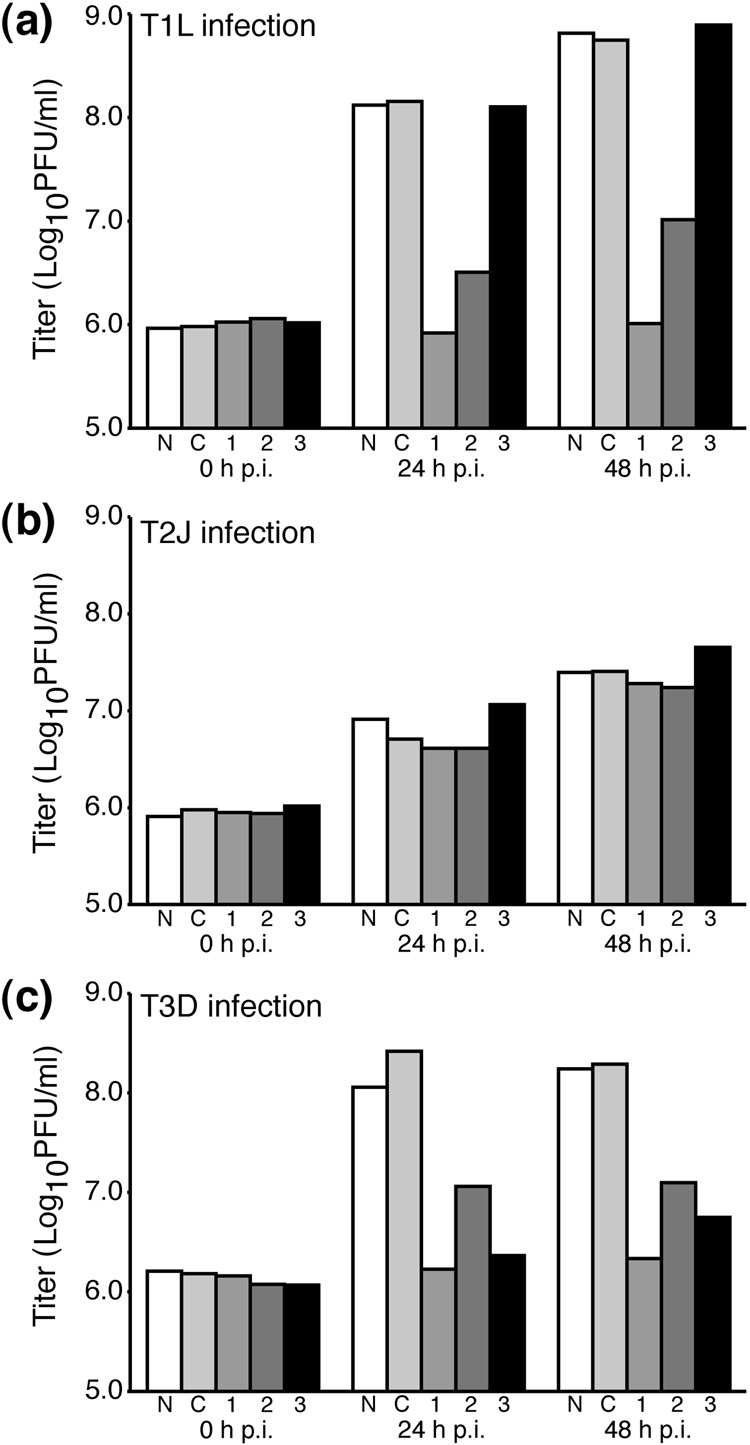
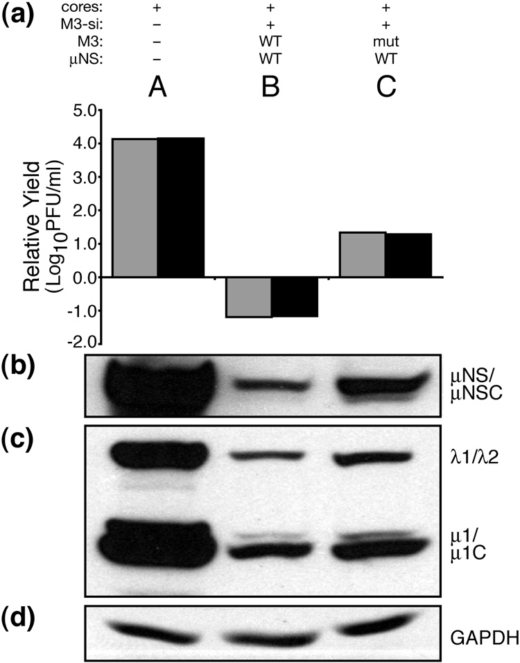
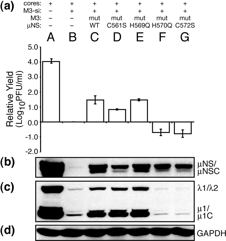
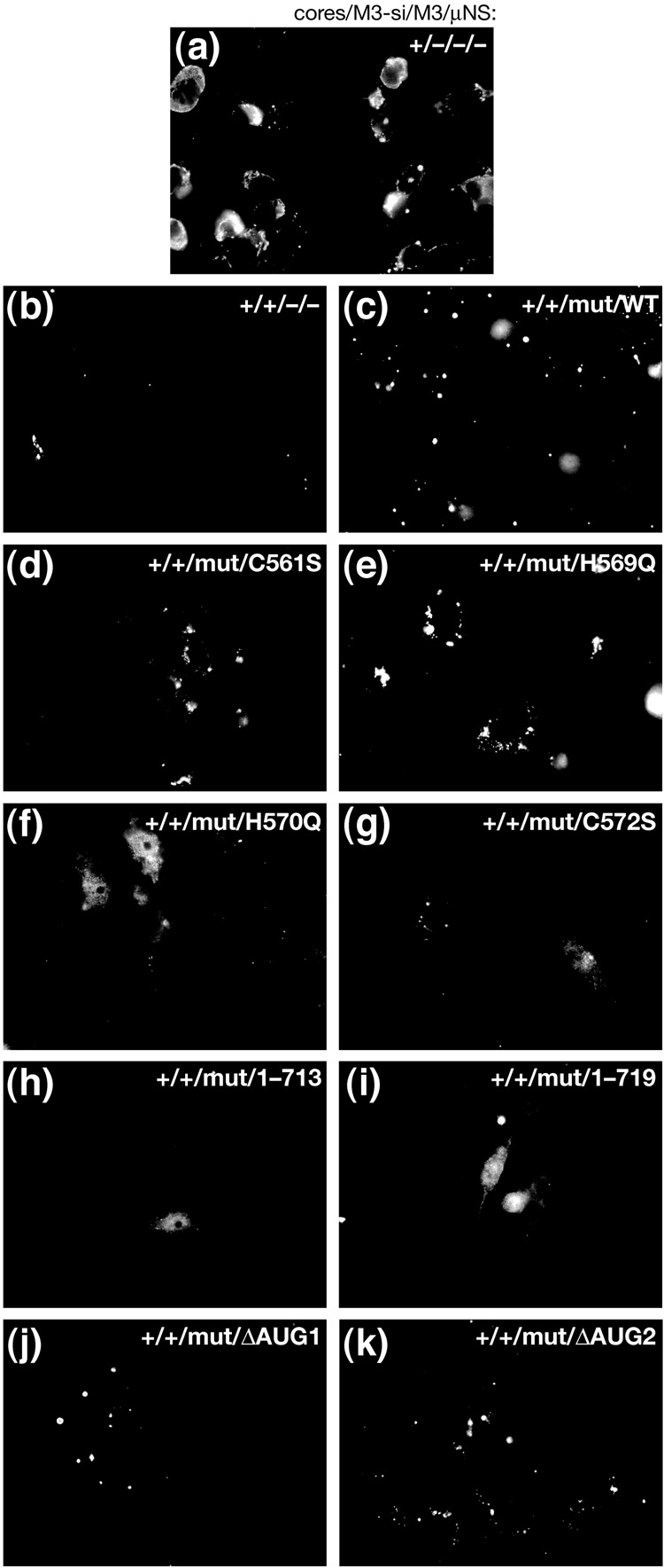
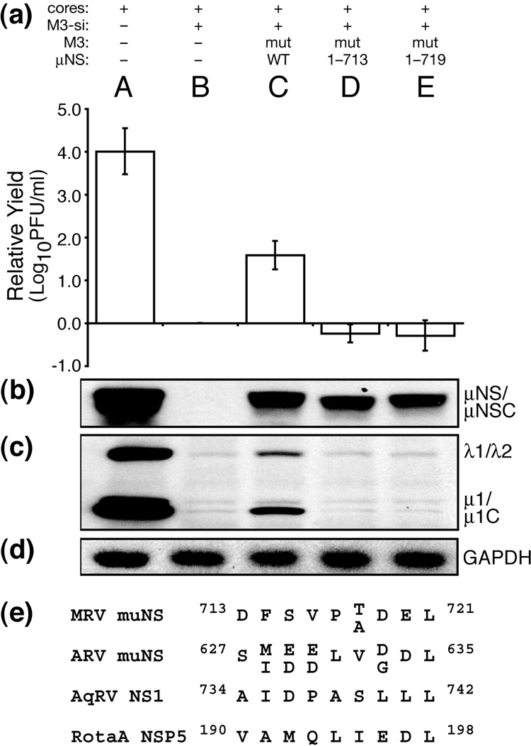
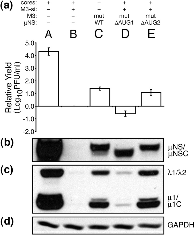
Genome replication of mammalian orthoreovirus (MRV) occurs in cytoplasmic inclusion bodies called viral factories. Nonstructural protein microNS, encoded by genome segment M3, is a major constituent of these structures. When expressed without other viral proteins, microNS forms cytoplasmic inclusions morphologically similar to factories, suggesting a role for microNS as the factory framework or matrix. In addition, most other MRV proteins, including all five core proteins (lambda1, lambda2, lambda3, micro2, and sigma2) and nonstructural protein sigmaNS, can associate with microNS in these structures. In the current study, small interfering RNA targeting M3 was transfected in association with MRV infection and shown to cause a substantial reduction in microNS expression as well as, among other effects, a reduction in infectious yields by as much as 4 log(10) values. By also transfecting in vitro-transcribed M3 plus-strand RNA containing silent mutations that render it resistant to the small interfering RNA, we were able to complement microNS expression and to rescue infectious yields by ~100-fold. We next used microNS mutants specifically defective at forming factory-matrix structures to show that this function of microNS is important for MRV growth; point mutations in a C-proximal, putative zinc-hook motif as well as small deletions at the extreme C terminus of microNS prevented rescue of viral growth while causing microNS to be diffusely distributed in cells. We furthermore confirmed that an N-terminally truncated form of microNS, designed to represent microNSC and still able to form factory-matrix structures, is unable to rescue MRV growth, localizing one or more other important functions to an N-terminal region of microNS known to be involved in both micro2 and sigmaNS association. Thus, factory-matrix formation is an important, though not a sufficient function of microNS during MRV infection; microNS is multifunctional in the course of viral growth.


Figures

Fig. 1
Fig. 1
siRNAs targeting MRV M3 transcripts. (a) Schematic of the M3 gene depicting locations of the siRNA target sequences in MRV strains T1L, T2J, and T3D. Nucleotides 194–212 of T1L and T3D are targeted by M3-si01 (1). Nucleotides 140–158 of T1L and T3D are targeted by M3-si02 (2). Nucleotides 1369–1387 of T3D are targeted by M3-si03 (3). (b) Sequences of the three M3-targeting siRNAs are shown, each followed by the corresponding region of the T1L, T2J, and T3D M3 genes. Mismatches between siRNA and gene are bolded and overlined in the gene sequence.
Fig. 2
Fig. 2
Viral protein expression and dsRNA production in siRNA-transfected cells. CV-1 cells were transfected by electroporation with either no siRNA, control siRNA, M3-si01, M3-si02, or M3-si03 as indicated in each panel. The cells were infected with MRV strain T1L, T2J, or T3D at 24 h p.t. at an MOI of 5. (a–c) Cells were harvested at 24 h p.i. by washing with PBS and then lysing in 2× sample buffer. Lysates were boiled for 5 min, separated by SDS/PAGE, transferred to nitrocellulose, and immunoblotted with polyclonal μNS antiserum (a) or polyclonal T1L virion antiserum (b). GAPDH immunoblot (c) was included as a loading control. (d) Cells were harvested at 24 h p.i. by washing with PBS and then purifying total RNA by TrizolLS extraction. Samples were heated to 60°C for 5 min, and dsRNAs (large, L; medium, M; and small, S as indicated) were resolved by SDS/PAGE. The gel was stained with 0.5% ethidium bromide.
Fig. 3
Fig. 3
Viral titers in siRNA-transfected cells. CV-1 cells were transfected by electroporation with either no siRNA (N), control siRNA (C), M3-si01 (1), M3-si02 (2), or M3-si03 (3) as indicated in each panel. The cells were infected with MRV strain T1L (a), T2J (b), or T3D (c) at 24 h p.t. at an MOI of 5. Cells were then harvested by freeze-thaw at the times specified in each panel, and infectious viral titers were determined by plaque assays on L929 cells. This figure is representative of three independent experiments.
Fig. 4
Fig. 4
Complementation of μNS with in vitro-transcribed M3 [+]RNA. BSRT7 cells were transfected using Lipofectamine 2000. Lanes reflect different combinations of transfected materials as follows: (A) T1L cores alone; (B) T1L cores, M3-si01, and in vitro-transcribed M3 [+]RNA expressing wild-type T1L μNS; and (C) T1L cores, M3-si01, and in vitro-transcribed M3 [+]RNA expressing wild-type T1L μNS but also containing silent mutations in the siRNA target sequence (mutRNA). Cells were then harvested at 24 h p.i. for viral titers and protein analyses. For titers (a), cells were harvested by freeze-thaw, and plaque assays were performed on L929 cells. Titers were expressed as infectious yields relative to that of the cores+M3-si01 sample (not shown in this figure) within each replicate experiment, and the relative yields from two such experiments are shown as adjacent bars for each type of sample. For protein analyses (b–d), cells were harvested by washing with PBS and then lysing in 2× sample buffer. Lysates were boiled for 5 min, separated by SDS/PAGE, transferred to nitrocellulose, and immunoblotted with polyclonal μNS antiserum (b) or T1L virion antiserum (c). GAPDH immunoblot (d) was included as a loading control. The blots are representative of the two experiments.
Fig. 5
Fig. 5
Rescue of viral growth with μNS mutants, part 1. BSRT7 cells were transfected using Lipofectamine 2000. Lanes reflect different combinations of transfected materials as follows: (A) T1L cores alone; (B) T1L cores and M3-si01; (C) T1L cores, M3-si01, and mutRNA expressing wild-type μNS as in Fig. 4; (D) T1L cores, M3-si01, and mutRNA with point mutaion C561S; (E) T1L cores, M3-si01, and mutRNA with point mutation H569Q; (F) T1L cores, M3-si01, and mutRNA with point mutation H570Q; and (G) T1L cores, M3-si01, and mutRNA with point mutation C572S. Cells were harvested at 24 h p.i. for viral titers (a) and protein analyses (b–d) as described for Fig. 4. Titers were expressed as infectious yields relative to that of the cores+M3-si01 sample within each replicate experiment, and the relative yields from three such experiments are shown as the mean ± standard deviation for each type of sample. The blots are representative of the three experiments.
Fig. 6
Fig. 6
Immunofluorescence microscopy of viral factories during rescue of viral growth with mutants of μNS. BSRT7 cells were transfected using Lipofectamine 2000 and then fixed at 24 h p.t. for immunostaining. The cells were immunostained with anti-μNS IgG followed by goat anti-rabbit IgG conjugated to Alexa 488. Images reflect different combinations of transfected materials as follows: (a) T1L cores alone; (b) T1L cores and M3-si01; (c) T1L cores, M3-si01, and mutRNA expressing wild-type μNS as in Fig. 4; (d) T1L cores, M3-si01, and mutRNA with point mutation C561S; (e) T1L cores, M3-si01, and mutRNA with point mutation H569Q; (f) T1L cores, M3-si01, and mutRNA with point mutation H570Q; (g) T1L cores, M3-si01, and mutRNA with point mutation C572S; (h) T1L cores, M3-si01, and mutRNA expressing μNS(1–713); (i) T1L cores, M3-si01, and mutRNA expressing μNS(1–719); (j) T1L cores, M3-si01, and mutRNA expressing μNS(41–721) (μNSC alone); and (k) T1L cores, M3-si01, and mutRNA expressing μNS(1–721) (μNS alone).
Fig. 7
Fig. 7
Rescue of viral growth with μNS mutants, part 2. (a–d) BSRT7 cells were transfected using Lipofectamine 2000. Lanes reflect different combinations of transfected materials as follows: (A) T1L cores alone; (B) T1L cores and M3-si01; (C) T1L cores, M3-si01, and mutRNA expressing wild-type μNS as in Fig. 4; (D) T1L cores, M3-si01, and mutRNA expressing μNS(1–713); and (E) T1L cores, M3-si01, and mutRNA expressing μNS(1–719). Cells were harvested at 24 h p.i. for viral titers (a) and protein analyses (b–d) as described for Fig. 4. Titers were expressed as relative yields as described for Fig. 5. The blots are representative of the three replicate experiments. (e) The C-terminal eight residues of μNS homologs from mammalian reoviruses (MRV), avian reoviruses (ARV), and aquareoviruses (AqRV NS1). Sequence variation between strains within each group of viruses is indicated by stacked letters. The C-terminal eight residues of viroplasm-forming protein NSP5 from group A rotavirus strains SA11 and RRV are also shown.
Fig. 8
Fig. 8
Expression of μNSC in the absence of μNS is unable to rescue viral growth. BSRT7 cells were transfected using Lipofectamine 2000. Lanes reflect different combinations of transfected materials as follows: (A) T1L cores alone; (B) T1L cores and M3-si01; (C) T1L cores, M3-si01, and mutRNA expressing wild-type μNS (and μNSC) as in Fig. 4; (D) T1L cores, M3-si01, and mutRNA expressing μNSC only (ΔAUG1); and (E) T1L cores, M3-si01, and mutRNA expressing full-length μNS only (ΔAUG2). Cells were harvested at 24 h p.i. for viral titers (a) and protein analyses (b–d) as described for Fig. 4. Titers were expressed as relative yields as described for Fig. 5. The blots are representative of the three replicate experiments.

References

    1. Ahmed R, Canning WM, Kauffman RS, Sharpe AH, Hallum JV, Fields BN. Role of the host cell in persistent viral infection: coevolution of L cells and reovirus during persistent infection. Cell. 1981;25:325–332. - PubMed
    1. Arias CF, Dector MA, Segovia L, Lopez T, Camacho M, Isa P, Espinosa R, Lopez S. RNA silencing of rotavirus gene expression. Virus Res. 2004;102:43–51. - PMC - PubMed
    1. Astell C, Silverstein SC, Levin DH, Acs G. Regulation of the reovirus RNA transcriptase by a viral capsomere protein. Virology. 1972;48:648–654. - PubMed
    1. Attoui H, Fang Q, Mohd Jaafar F, Cantaloube JF, Biagini P, de Micco P, de Lamballerie X. Common evolutionary origin of aquareoviruses and orthoreoviruses revealed by genome characterization of Golden shiner reovirus, Grass carp reovirus, Striped bass reovirus and golden ide reovirus (genus Aquareovirus, family Reoviridae) J. Gen. Virol. 2002;83:1941–1951. - PubMed
    1. Banerjee AK, Shatkin AJ. Transcription in vitro by reovirus-associated ribonucleic acid-dependent polymerase. J. Virol. 1970;6:1–11. - PMC - PubMed

Publication types

MeSH terms

Substances