

Translocator protein 18 kDa (TSPO): molecular sensor of brain injury and repair
- PMID: 18374421
- PMCID: PMC2453598
- DOI: 10.1016/j.pharmthera.2007.12.004
Translocator protein 18 kDa (TSPO): molecular sensor of brain injury and repair
Abstract
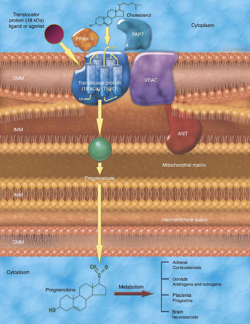
For over 15 years, the peripheral benzodiazepine receptor (PBR), recently named translocator protein 18 kDa (TSPO) has been studied as a biomarker of reactive gliosis and inflammation associated with a variety of neuropathological conditions. Early studies documented that in the brain parenchyma, TSPO is exclusively localized in glial cells. Under normal physiological conditions, TSPO levels are low in the brain neuropil but they markedly increase at sites of brain injury and inflammation making it uniquely suited for assessing active gliosis. This research has generated significant efforts from multiple research groups throughout the world to apply TSPO as a marker of "active" brain pathology using in vivo imaging modalities such as Positron Emission Tomography (PET) in experimental animals and humans. Further, in the last few years, there has been an increased interest in understanding the molecular and cellular function(s) of TSPO in glial cells. The latest evidence suggests that TSPO may not only serve as a biomarker of active brain disease but also the use of TSPO-specific ligands may have therapeutic implications in brain injury and repair. This review presents an overview of the history and function of TSPO focusing on studies related to its use as a sensor of active brain disease in experimental animals and in human studies.
Figures
References
-
- Alho H, Harjuntausta T, Schultz R, Pelto-Huikko M, Bovolin P. Immunohistochemistry of diazepam binding inhibitor (DBI) in the central nervous system and peripheral organs: its possible role as an endogenous regulator of different types of benzodiazepine receptors. Neuropharmacology. 1991;30:1381–1386. - PubMed
-
- Alho H, Varga V, Krueger KE. Expression of mitochondrial benzodiazepine receptor and its putative endogenous ligand diazepam binding inhibitor in cultured primary astrocytes and C-6 cells: relation to cell growth. Cell Growth Differ. 1994;5:1005–1014. - PubMed
-
- Altar CA, Baudry M. Systemic injection of kainic acid: gliosis in olfactory and limbic brain regions quantified with [3H]-PK 11195 binding autoradiography. Exp Neurol. 1990;109:333–341. - PubMed
-
- Anholt RR, Pedersen PL, De Souza EB, Snyder SH. The peripheral-type benzodiazepine receptor. Localization to the mitochondrial outer membrane. J Biol Chem. 1986;261:576–583. - PubMed
-
- Antkiewicz-Michaluk L, Guidotti A, Krueger KE. Molecular characterization and mitochondrial density of a recognition site for peripheral-type benzodiazepine ligands. Mol Pharmacol. 1988;34:272–278. - PubMed
Publication types
MeSH terms
Substances
Grants and funding
LinkOut - more resources
Full Text Sources
Other Literature Sources
