Comparative Study
. 2008 May;38(1):80-8.
doi: 10.1016/j.mcn.2008.01.017. Epub 2008 Feb 20.

Schwann cell proliferation during Wallerian degeneration is not necessary for regeneration and remyelination of the peripheral nerves: axon-dependent removal of newly generated Schwann cells by apoptosis

Affiliations
Comparative Study

Schwann cell proliferation during Wallerian degeneration is not necessary for regeneration and remyelination of the peripheral nerves: axon-dependent removal of newly generated Schwann cells by apoptosis

David P Yang et al. Mol Cell Neurosci. 2008 May.

Abstract

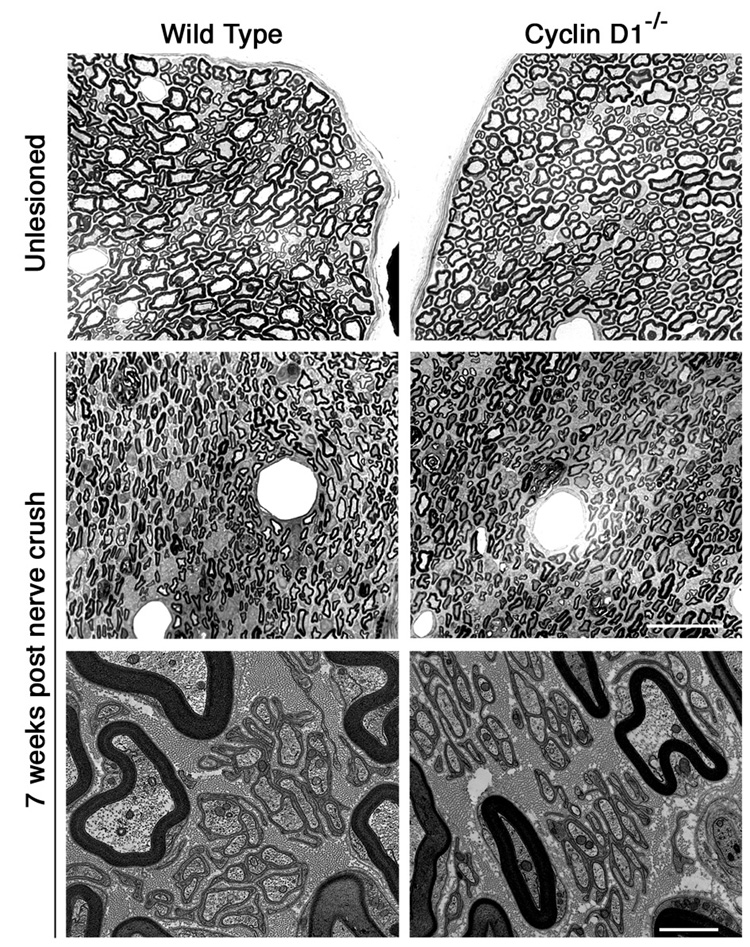
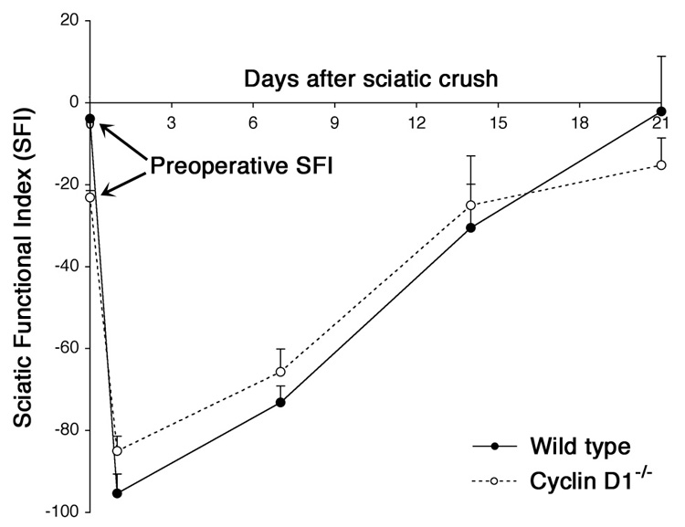
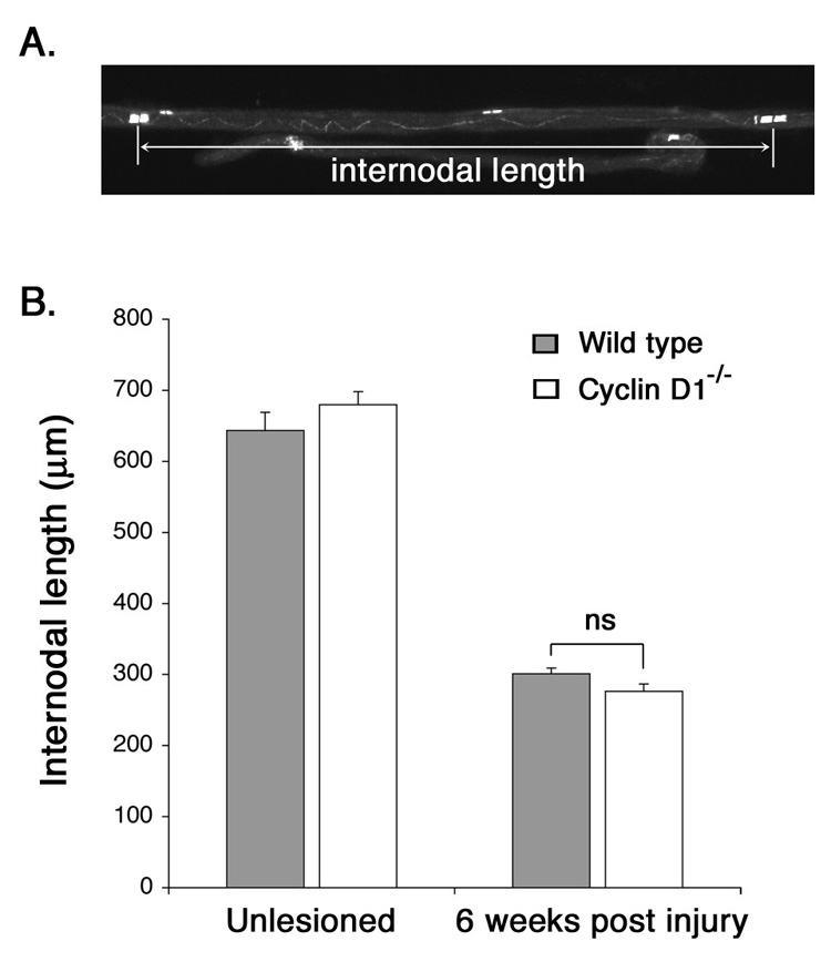
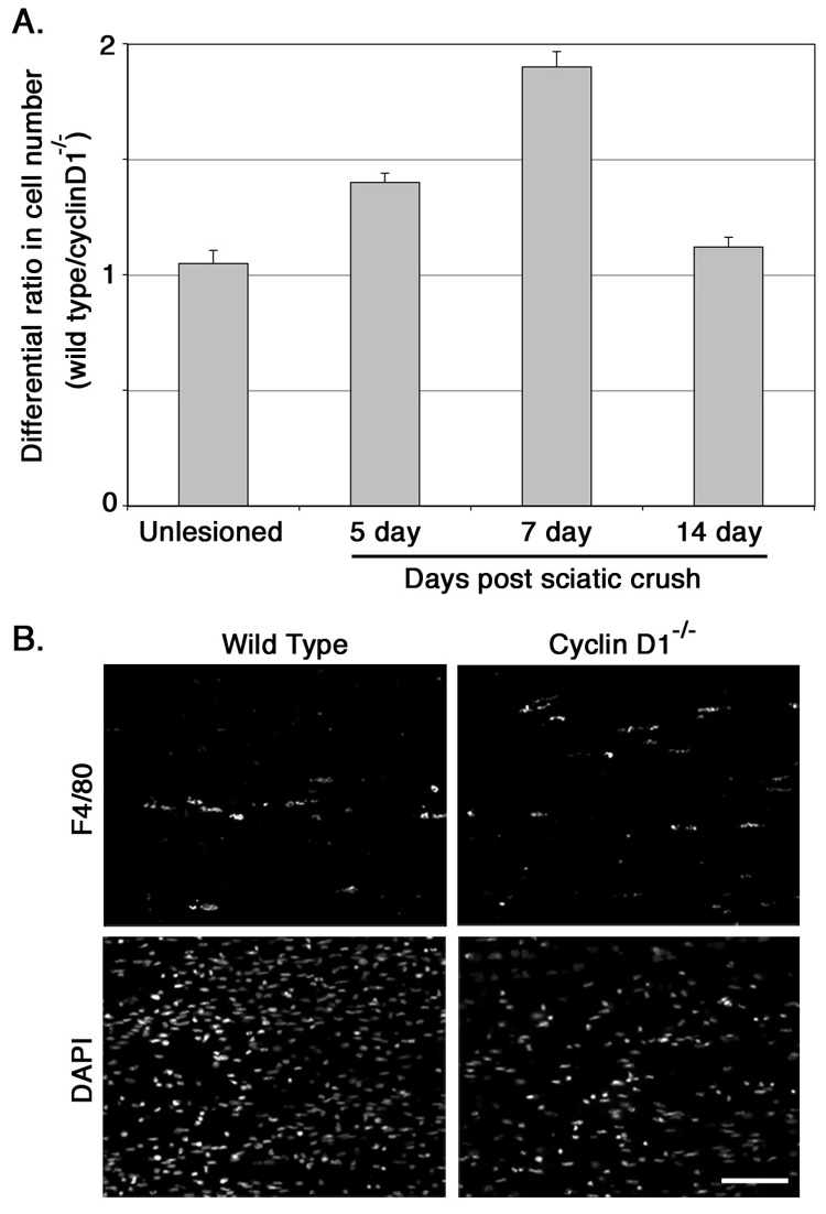
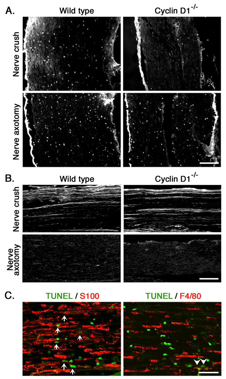
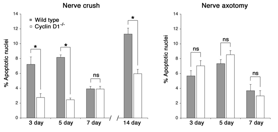
Peripheral nerve injury is followed by a wave of Schwann cell proliferation in the distal nerve stumps. To resolve the role of Schwann cell proliferation during functional recovery of the injured nerves, we used a mouse model in which injury-induced Schwann cell mitotic response is ablated via targeted disruption of cyclin D1. In the absence of distal Schwann cell proliferation, axonal regeneration and myelination occur normally in the mutant mice and functional recovery of injured nerves is achieved. This is enabled by pre-existing Schwann cells in the distal stump that persist but do not divide. On the other hand, in the wild type littermates, newly generated Schwann cells of injured nerves are culled by apoptosis. As a result, distal Schwann cell numbers in wild type and cyclin D1 null mice converge to equivalence in regenerated nerves. Therefore, distal Schwann cell proliferation is not required for functional recovery of injured nerves.


Figures

Figure 1
Figure 1
Morphology of the distal sciatic nerves of wild type and cyclin D1−/− mice. Top panels: Semi-thin transverse sections of unlesioned sciatic nerves of wild type and cyclin D1−/− mice. Middle panels: Seven weeks following sciatic nerve crush. Regenerated axons are smaller in diameter than axons in the unlesioned nerve. Axonal density and morphology of cyclin D1−/− nerve do not appear to be different from wild type nerve. Scale bar: 60 µm. Bottom panels: Ultra-thin transverse sections are shown in order to visualize myelinated and non-myelinated axons of the regenerated sciatic nerve. Large axons are myelinated normally and small axons within Remak bundles are well segregated by Schwann cell plasma membrane in both groups. Scale bar: 20 µm
Figure 2
Figure 2
Sciatic nerve function recovers normally in the absence of distal Schwann cell proliferation in cyclin D1−/− mice. Sciatic nerve function was evaluated by determining Sciatic Function Index (SFI) from parameters obtained from walking track analysis before (preoperative SFI) and after sciatic nerve crush. Soon after nerve crush SFI in both animals decreased to levels near −100, representing complete loss of function. The SFI recovered to the pre-operative values by 3 weeks in both groups. No significant difference in the recovery rate was observed between wild type and cyclin D1−/− mice. The means (±SEM) were calculated from four animals per group.
Figure 3
Figure 3
Myelin segments on regenerated axons are similar in length in wild type and cyclin D1−/− mice. A. Length of a myelin segment was determined by measuring the internodal length. Teased nerve fibers were prepared and immunostained for a paranodal protein Caspr. Internodal length is determined by a distance between two nodes along an axon, with each node encompassed by Caspr+ paranodes. B. Comparison of average myelin segment lengths associated with unlesioned or regenerated axons (6 weeks following sciatic nerve crush) between wild type and cyclin D1−/− mice. The difference between the two groups is not statistically significant (ns). The means (±SEM) were calculated from analyzing 80–120 myelin segments from three animals per group. Statistical significance was determined by ANOVA test.
Figure 4
Figure 4
Difference in distal cell number between wild type and cyclin D1−/− mice diminishes as the regeneration progresses. A. Nerve sections were prepared from unlesioned sciatic nerve or distal nerve at 5, 7 and 14 days following nerve crush. The cell nuclei were visualized by DAPI-labeling and counted. The data represents ratio between distal cell number from wild type and cyclin D1−/− mice. In unlesioned nerve, the ratio is close to 1, indicating indifference in distal cell density between the two groups. Following nerve crush, differences in cell density widen reflecting Schwann cell proliferation in wild type but not in cyclin D1−/− distal nerve. At 14 days, the ratio decreases to a value close to 1. The data represent the mean ratio (±SEM) determined from 50 sets of wild type and cyclin D1−/− fields obtained from three animals per group. B. Seven-day distal nerve sections were immunostained for F4/80 to visualize macrophages. No apparent difference in macrophage number is seen between wild type and cyclin D1−/− mice despite the increase in distal cell number (DAPI+) in the wild type.
Figure 5
Figure 5
Excess Schwann cells generated in wild type mice are removed by apoptosis. A. Longitudinal sections of distal sciatic nerve five days after nerve crush and axotomy. Apoptotic cells are labeled by TUNEL assay. A higher percentage of TUNEL+ apoptotic cells are detected in wild type distal nerve following nerve crush and axotomy. Very few apoptotic cells are seen in cyclin D1−/− section after nerve crush. However, this proportion increases drastically when axonal regeneration is prevented by axotomy. Scale bar: 80 µm. B. The distal nerve sections were also immunostained for GAP-43 to visualize growing axons. Regenerating axons are clearly seen in distal nerve section from wild type and cyclic D1−/− mice 5 days after nerve crush but not after nerve axotomy. Scale bar: 70 µm. C. Confocal images of distal nerve sections double-immunostained for apoptotic cells (green) and S100 or macrophage specific F4/80 antigen (red). Most of the apoptotic cells express S100 (arrows, left panel), indicating that they are Schwann cells. Most of the apoptotic cells did not express F4/80 (right panel). Occasionally, few apoptotic macrophages were observed (arrow head). Scale bar: 30 µm.
Figure 6
Figure 6
Survival of distal denervated Schwann cells is dependent on the presence of regenerating axons. Distal nerve sections from wild type and cyclin D1−/− sciatic nerves prepared at 3, 5, 7 and 14 days following nerve crush, or 3, 5, 7 days following nerve axotomy were processed for detection of apoptosis by TUNEL assay. The data represent the mean percentage of apoptotic cells (± SEM) in 30–50 fields obtained from three animals. Statistical significance was determined by ANOVA test. *: p < 0.0001, ns: not significant

Similar articles

Cited by

References

    1. Aldskogius H, Molander C, Persson J, Thomander L. Specific and nonspecific regeneration of motor axons after sciatic nerve injury and repair in the rat. J Neurol Sci. 1987;80:249–257. - PubMed
    1. Araki T, Milbrandt J. Ninjurin, a novel adhesion molecule, is induced by nerve injury and promotes axonal growth. Neuron. 1996;17:353–361. - PubMed
    1. Atanasoski S, Shumas S, Dickson C, Scherer SS, Suter U. Differential cyclin D1 requirements of proliferating Schwann cells during development and after injury. Mol Cell Neurosci. 2001;18:581–592. - PubMed
    1. Banner LR, Patterson PH. Major changes in the expression of the mRNAs for cholinergic differentiation factor/leukemia inhibitory factor and its receptor after injury to adult peripheral nerves and ganglia. Proc Natl Acad Sci U S A. 1994;91:7109–7113. - PMC - PubMed
    1. Barrett GL, Bartlett PF. The p75 nerve growth factor receptor mediates survival or death depending on the stage of sensory neuron development. Proc Natl Acad Sci U S A. 1994;91:6501–6505. - PMC - PubMed

Publication types

MeSH terms