Ibuprofen reduces Abeta, hyperphosphorylated tau and memory deficits in Alzheimer mice
- PMID: 18374906
- PMCID: PMC2587244
- DOI: 10.1016/j.brainres.2008.01.095
Ibuprofen reduces Abeta, hyperphosphorylated tau and memory deficits in Alzheimer mice
Abstract
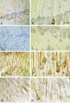
We examined the effects of ibuprofen on cognitive deficits, Abeta and tau accumulation in young triple transgenic (3xTg-AD) mice. 3xTg-AD mice were fed ibuprofen-supplemented chow between 1 and 6 months. Untreated 3xTg-AD mice showed significant impairment in the ability to learn the Morris water maze (MWM) task compared to age-matched wild-type (WT) mice. The performance of 3xTg-AD mice was significantly improved with ibuprofen treatment compared to untreated 3xTg-AD mice. Ibuprofen-treated transgenic mice showed a significant decrease in intraneuronal oligomeric Abeta and hyperphosphorylated tau (AT8) immunoreactivity in the hippocampus. Confocal microscopy demonstrated co-localization of conformationally altered (MC1) and early phosphorylated tau (CP-13) with oligomeric Abeta, and less co-localization of oligomeric Abeta and later forms of phosphorylated tau (AT8 and PHF-1) in untreated 3xTg-AD mice. Our findings show that prophylactic treatment of young 3xTg-AD mice with ibuprofen reduces intraneuronal oligomeric Abeta, reduces cognitive deficits, and prevents hyperphosphorylated tau immunoreactivity. These findings provide further support for intraneuronal Abeta as a cause of cognitive impairment, and suggest that pathological alterations of tau are associated with intraneuronal oligomeric Abeta accumulation.
Figures


Similar articles
-
Ultrasound with microbubbles improves memory, ameliorates pathology and modulates hippocampal proteomic changes in a triple transgenic mouse model of Alzheimer's disease.Theranostics. 2020 Sep 26;10(25):11794-11819. doi: 10.7150/thno.44152. eCollection 2020. Theranostics. 2020. PMID: 33052247 Free PMC article.
-
Immunotherapy to improve cognition and reduce pathological species in an Alzheimer's disease mouse model.Alzheimers Res Ther. 2018 Jun 18;10(1):54. doi: 10.1186/s13195-018-0384-9. Alzheimers Res Ther. 2018. PMID: 29914551 Free PMC article.
-
Fyn knock-down increases Aβ, decreases phospho-tau, and worsens spatial learning in 3×Tg-AD mice.Neurobiol Aging. 2012 Apr;33(4):825.e15-24. doi: 10.1016/j.neurobiolaging.2011.05.014. Epub 2011 Jul 7. Neurobiol Aging. 2012. PMID: 21741124 Free PMC article.
-
Cognitive Decline in Preclinical Alzheimer's Disease: Amyloid-Beta versus Tauopathy.J Alzheimers Dis. 2018;61(1):265-281. doi: 10.3233/JAD-170490. J Alzheimers Dis. 2018. PMID: 29154274 Free PMC article. Review.
-
Effects of CX3CR1 and Fractalkine Chemokines in Amyloid Beta Clearance and p-Tau Accumulation in Alzheimer's Disease (AD) Rodent Models: Is Fractalkine a Systemic Biomarker for AD?Curr Alzheimer Res. 2016;13(4):403-12. doi: 10.2174/1567205013666151116125714. Curr Alzheimer Res. 2016. PMID: 26567742 Review.
Cited by
-
Modulation of Aβ42 in vivo by γ-secretase modulator in primates and humans.Alzheimers Res Ther. 2015 Aug 5;7(1):55. doi: 10.1186/s13195-015-0137-y. eCollection 2015. Alzheimers Res Ther. 2015. PMID: 26244059 Free PMC article.
-
Disease modifying drugs targeting β-amyloid.Am J Alzheimers Dis Other Demen. 2012 Aug;27(5):296-300. doi: 10.1177/1533317512452034. Am J Alzheimers Dis Other Demen. 2012. PMID: 22815077 Free PMC article. Review.
-
Lysine reactivity profiling reveals molecular insights into human serum albumin-small-molecule drug interactions.Anal Bioanal Chem. 2021 Dec;413(30):7431-7440. doi: 10.1007/s00216-021-03700-1. Epub 2021 Oct 21. Anal Bioanal Chem. 2021. PMID: 34676431
-
The γ-secretase modulator CHF5074 reduces the accumulation of native hyperphosphorylated tau in a transgenic mouse model of Alzheimer's disease.J Mol Neurosci. 2011 Sep;45(1):22-31. doi: 10.1007/s12031-010-9482-2. Epub 2010 Dec 22. J Mol Neurosci. 2011. PMID: 21181298
-
The effects of aging, housing and ibuprofen treatment on brain neurochemistry in a triple transgene Alzheimer's disease mouse model using magnetic resonance spectroscopy and imaging.Brain Res. 2014 Nov 24;1590:85-96. doi: 10.1016/j.brainres.2014.09.067. Epub 2014 Oct 6. Brain Res. 2014. PMID: 25301691 Free PMC article.
References
-
- Agdeppa ED, Kepe V, Petri A, Satyamurthy N, Liu J, Huang SC, Small GW, Cole GM, Barrio JR. In vitro detection of (S)-naproxen and ibuprofen binding to plaques in the Alzheimer's brain using the positron emission tomography molecular imaging probe 2-(1-[6-[(2-[(18)F]fluoroethyl)(methyl)amino]-2-naphthyl]ethylidene)malono nitrile. Neuroscience. 2003;117:723–730. - PubMed
-
- Akiyama H, Barger S, Barnum S, Bradt B, Bauer J, Cole GM, Cooper NR, Eikelenboom P, Emmerling M, Fiebich BL, Finch CE, Frautschy S, Griffin WS, Hampel H, Hull M, Landreth G, Lue L, Mrak R, Mackenzie IR, McGeer PL, O'Banion MK, Pachter J, Pasinetti G, Plata-Salaman C, Rogers J, Rydel R, Shen Y, Streit W, Strohmeyer R, Tooyoma I, Van Muiswinkel FL, Veerhuis R, Walker D, Webster S, Wegrzyniak B, Wenk G, Wyss-Coray T. Inflammation and Alzheimer's disease. Neurobiol Aging. 2000;21:383–421. - PMC - PubMed
-
- Barghorn S, Mandelkow E. Toward a unified scheme for the aggregation of tau into Alzheimer paired helical filaments. Biochemistry. 2002;41:14885–14896. - PubMed
-
- Billings LM, Oddo S, Green KN, McGaugh JL, LaFerla FM. Intraneuronal Abeta causes the onset of early Alzheimer's disease-related cognitive deficits in transgenic mice. Neuron. 2005;45:675–688. - PubMed
Publication types
MeSH terms
Substances
Grants and funding
LinkOut - more resources
Full Text Sources
Other Literature Sources
Medical
Molecular Biology Databases
Miscellaneous

