

Monolithic porous polymer stationary phases in polyimide chips for the fast high-performance liquid chromatography separation of proteins and peptides
- PMID: 18374934
- PMCID: PMC2518059
- DOI: 10.1016/j.chroma.2008.03.025
Monolithic porous polymer stationary phases in polyimide chips for the fast high-performance liquid chromatography separation of proteins and peptides
Abstract
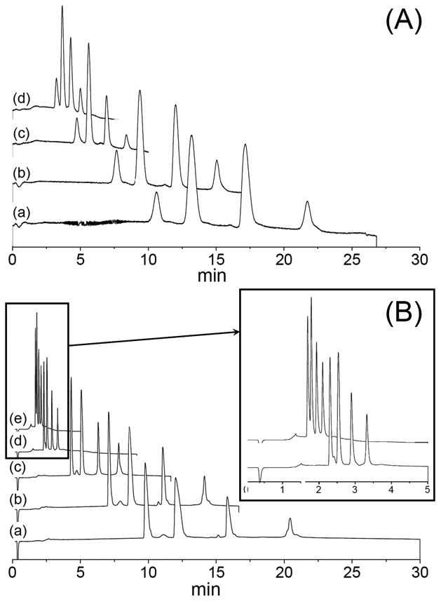
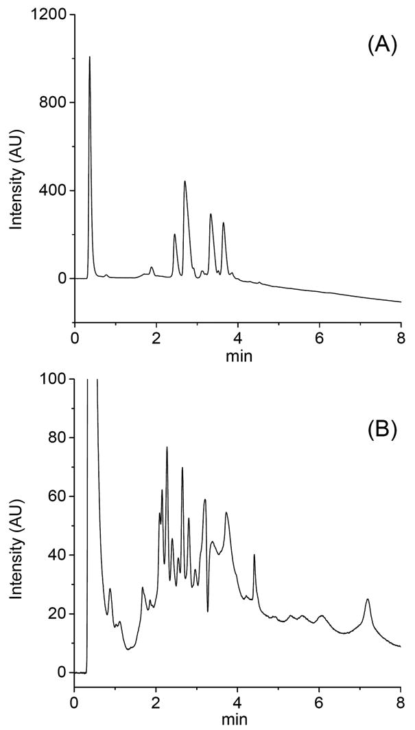
Poly(lauryl methacrylate-co-ethylene dimethacrylate) and poly(styrene-co-divinylbenzene) stationary phases in monolithic format have been prepared by thermally initiated free radical polymerization within polyimide chips featuring channels having a cross-section of 200micromx200microm and a length of 6.8cm. These chips were then used for the separation of a mixture of proteins including ribonuclease A, myoglobin, cytochrome c, and ovalbumin, as well as peptides. The separations were monitored by UV adsorption. Both the monolithic phases based on methacrylate and on styrene chemistries enabled the rapid baseline separation of most of the test mixtures. Best performance was achieved with the styrenic monolith leading to fast baseline separation of all four proteins in less than 2.5min. The in situ monolith preparation process affords microfluidic devices exhibiting good batch-to-batch and injection-to-injection repeatability.
Figures


Similar articles
-
Polymerisation and surface modification of methacrylate monoliths in polyimide channels and polyimide coated capillaries using 660 nm light emitting diodes.J Chromatogr A. 2011 May 20;1218(20):2954-62. doi: 10.1016/j.chroma.2011.03.021. Epub 2011 Mar 21. J Chromatogr A. 2011. PMID: 21477803
-
Rapid reversed-phase separation of proteins and peptides using optimized 'moulded' monolithic poly(styrene-co-divinylbenzene) columns.J Chromatogr A. 1999 Dec 31;865(1-2):169-74. doi: 10.1016/s0021-9673(99)00981-4. J Chromatogr A. 1999. PMID: 10674939
-
Organic polymer monoliths as stationary phases for capillary HPLC.J Sep Sci. 2004 Dec;27(17-18):1419-30. doi: 10.1002/jssc.200401825. J Sep Sci. 2004. PMID: 15638150 Review.
-
Effect of capillary cross-section geometry and size on the separation of proteins in gradient mode using monolithic poly(butyl methacrylate-co-ethylene dimethacrylate) columns.J Chromatogr A. 2009 Mar 20;1216(12):2355-61. doi: 10.1016/j.chroma.2009.01.007. Epub 2009 Jan 9. J Chromatogr A. 2009. PMID: 19201413 Free PMC article.
-
Preparation and characterization of methacrylate-based semi-micro monoliths for high-throughput bioanalysis.Anal Bioanal Chem. 2006 Oct;386(3):566-71. doi: 10.1007/s00216-006-0425-2. Epub 2006 May 10. Anal Bioanal Chem. 2006. PMID: 16685518 Review.
Cited by
-
Polymer microchips integrating solid-phase extraction and high-performance liquid chromatography using reversed-phase polymethacrylate monoliths.Anal Chem. 2009 Apr 1;81(7):2545-54. doi: 10.1021/ac802359e. Anal Chem. 2009. PMID: 19267447 Free PMC article.
-
Use of photopatterned porous polymer monoliths as passive micromixers to enhance mixing efficiency for on-chip labeling reactions.Lab Chip. 2009 Apr 7;9(7):877-83. doi: 10.1039/b816521a. Epub 2009 Jan 7. Lab Chip. 2009. PMID: 19294297 Free PMC article.
-
Visible light initiated polymerization of styrenic monolithic stationary phases using 470 nm light emitting diode arrays.J Sep Sci. 2010 Jan;33(1):61-6. doi: 10.1002/jssc.200900624. J Sep Sci. 2010. PMID: 20091717 Free PMC article.
-
Advances in monoliths and related porous materials for microfluidics.Biomicrofluidics. 2016 May 4;10(3):032901. doi: 10.1063/1.4948507. eCollection 2016 May. Biomicrofluidics. 2016. PMID: 27190564 Free PMC article. Review.
-
Recent advances in lab-on-a-chip technologies for viral diagnosis.Biosens Bioelectron. 2020 Apr 1;153:112041. doi: 10.1016/j.bios.2020.112041. Epub 2020 Jan 22. Biosens Bioelectron. 2020. PMID: 31999560 Free PMC article. Review.
References
-
- Hamdan M, Righetti PG. Proteomics Today. Wiley; Hoboken, NJ: 2005.
-
- Cooper JW, Wang Y, Lee CS. Electrophoresis. 2004;25:3913. - PubMed
-
- Lion N, Rohner TC, Dayon L, Arnaud IL, Damoc E, Youhnovski N, Wu Z, Roussel C, Josserand J, Jensen H, Rossier JS, Przybylski M, Girault HH. Electrophoresis. 2003;24:3533. - PubMed
-
- Hardouin J, Duchateau M, Joubert-Caron R, Caron M. Rapid Commun Mass Spectrom. 2006;20:3236. - PubMed
-
- Ocvirk G, Verpoorte E, Manz A, Grasserbauer M, Widmer HM. Anal Methods Instrum. 1995;2:74.
Publication types
MeSH terms
Substances
Grants and funding
LinkOut - more resources
Full Text Sources
Other Literature Sources
