. 2008 Jun 15;118(1):13-20.
doi: 10.1016/j.imlet.2008.02.005. Epub 2008 Mar 13.

Stimulation of T cells up-regulates expression of Ifi202, an interferon-inducible lupus susceptibility gene, through activation of JNK/c-Jun pathway

Affiliations

Stimulation of T cells up-regulates expression of Ifi202, an interferon-inducible lupus susceptibility gene, through activation of JNK/c-Jun pathway

Jianming Chen et al. Immunol Lett. .

Abstract

Studies have revealed that increased expression of interferon (IFN)-inducible Ifi202 gene (encoding p202 protein) in splenic B and T cells from B6.Nba2 congenic (congenic for Nb2 locus derived from NZB mice) female mice is associated with lupus susceptibility. However, signaling pathways that regulate Ifi202 expression in immune cells remain to be elucidated. Here we report that stimulation of T cells up-regulates the Ifi202 expression. We found that steady-state levels of Ifi202 mRNA and protein were detectable in splenic T cells from NZB mice and stimulation of T cells with anti-CD3 and anti-CD28 up-regulated expression of the Ifi202 gene. Similarly, stimulation of cells of a mouse T cell hybridoma cell line (2B4.11) also activated transcription of the Ifi202 gene. Significantly, up-regulation of Ifi202 expression in stimulated T cells was inhibited by treatment of cells with SP600125, a specific inhibitor of c-Jun N-terminal kinase (JNK). Conversely, treatment of cells with anisomycin, a potent activator of the JNK and c-Jun, up-regulated Ifi202 expression. Consistent with the activation of JNK/c-Jun pathway by T cell stimulation, forced expression of c-Jun in 2B4 T cells and in mouse embryonic fibroblasts (MEFs) also up-regulated the Ifi202 expression. Furthermore, we found that stimulation of T cells increased association of the activated c-Jun to the 5'-regulatory region of the Ifi202 gene in chromatin immunoprecipitation assays (ChIPs). Together, our observations demonstrate that stimulation of T cells up-regulates the Ifi202 expression in part through the JNK/c-Jun pathway.


Figures

Fig. 1
Fig. 1. Expression of Ifi202 is detectable in splenic purified T cells from NZB mice
(A) Total RNA was isolated from freshly prepared purified splenic T cells (lane 3) or splenic cells depleted of T cells (lane 4) as described in methods. The isolated RNA was subjected to semi-quantitative RT-PCR using a pair of primers specific to the Ifi202 gene or actin. As a control for Ifi202 expression, total RNA from mouse L929 cells, either left untreated (lane 1) or treated with IFN-α (1,000 u/ml for 24 h) (lane 2), was also subjected to RT-PCR. (B) Total cell extracts prepared from purified splenic T- (lane 3) or B-cells (lane 4) were subjected to immunoblotting using antibodies specific to the indicated proteins. As a negative and a positive control for immunoblotting, total cell extracts from mouse L929 cells, either left untreated (lane 1) or treated with IFN-α (1,000 u/ml for 24 h) (lane 2), were also subjected to immunoblotting.
Fig. 2
Fig. 2. Stimulation of splenic T-cells up-regulates Ifi202 expression
(A) Freshly isolated splenocytes (2–4 x10⁶) from age-matched (~ 12-weeks) male (lanes 1 and 2) or female (lanes 3 and 4) mice were plated in 60 mm plastic cell culture plates either coated with purified hamster anti-mouse CD3 epsilon (10 μg/plate) antibody or purified golden Syrian hamster IgG (5 μg/plate) isotype control antibody. Purified anti-mouse CD28 antibody (2 μg/ml) was added to the medium immediately after plating of splenocytes. After incubation of cells for 24 h, cells were collected and processed for isolation of total RNA. Isolated RNA was subjected to semi-quantitative RT-PCR using a pair of primers specific to Ifi202 gene or actin. (B) Freshly isolated splenocytes (2–4 x10⁶) from age-matched male (lanes 3 and 4) or female (lanes 5 and 6) mice were subjected to stimulation of T cells for 24 h as described in (A) above and cells were processed for isolation of total proteins. Total cell lysates were subjected to immunoblotting using antibodies specific to the indicated proteins. As a control, we also analyzed extracts from untreated (lane 1) or IFN-alpha treated (lane 2) L929 cells.
Fig. 3
Fig. 3. Stimulation of 2B4 cells up-regulates the expression of Ifi202 gene
(A) The mouse 2B4 cells (2–4 x10⁶) were incubated with anti-mouse CD3 epsilon (10 μg/plate) and anti-mouse CD28 antibody (2 μg/ml) for either 0 (lane 1), 0.5 (lane 2), 3 (lane 3), 6 (lane 4), 12 (lane 5), 24 (lane 6), or 36 h (lane 7) for stimulation. After the stimulation, cells were collected and processed for isolation of total RNA. The RNA was subjected to semi-quantitative RT-PCR using a pair of primers specific to Ifi202 gene or actin. (B) 2B4 cells (2–4 x10⁶) were stimulated with anti-mouse CD3 epsilon (10 μg/plate) and anti-mouse CD28 antibody (2 μg/ml) for either 0 (lane 1), 0.5 (lane 2), 6 (lane 3), 12 (lane 4), 24 (lane 5), or 36 h (lane 6). After the stimulation, cells were collected and lysed. Total cell lysates were subjected to immunoblotting using antibodies specific to the indicated proteins.
Fig. 4
Fig. 4. Up-regulation of the Ifi202 expression after T cell stimulation depends on activation of the JNK/c-Jun pathway
(A) 2B4 cells (2–4 x10⁶) were incubated with either an isotype control antibody (lane 1) or stimulated with anti-mouse CD3 epsilon (10 μg/plate) and anti-mouse CD28 antibody (2 μg/ml) for 0.5 (lane 2), 6 (lane 3), 12 (lane 4), 24 (lane 5), or 36 h (lane 6). After stimulation of cells, cells were collected and lysed. Total cell lysates were subjected to immunoblotting using antibodies specific to the indicated proteins. (B) 2B4 cells (2–4 x10⁶) were incubated with either an isotype control antibody (lane 1 and 4) or stimulated with anti-mouse CD3 epsilon (10 μg/plate) and anti-mouse CD28 antibody (2 μg/ml) for 12 h (lanes 2 and 5) or 24 h (lanes 3 and 6) in the absence (lanes 1–3) or presence (lanes 4–6) JNK inhibitor SP600125 (20 μM). After incubations, cells were collected and processed for isolation of total RNA. The RNA was subjected to semi-quantitative RT-PCR using a pair of primers specific to Ifi202 gene or actin. (C) 2B4 cells (2–4 x10⁶) were incubated with either an isotype control antibody (lane 1 and 4) or stimulated with anti-mouse CD3 epsilon (10 μg/plate) and anti-mouse CD28 antibody (2 μg/ml) for 12 h (lanes 2 and 5) or 24 h (lanes 3 and 6) in the absence (lanes 1–3) or presence (lanes 4–6) of JNK inhibitor SP600125 (10 μM). After incubations, cells were lysed. Total cell lysates were subjected to immunoblotting using antibodies specific to the indicated proteins. (D) 2B4 cells (2–4 x10⁶) were incubated without any treatment (lane 1) or, as a control, with DMSO (lane 2), or anisomycin (20 μM in DMSO) for the indicated length of time. After incubations, cells were lysed. Total cell lysates were subjected to immunoblotting using antibodies specific to the indicated proteins. (E) Freshly isolated splenocytes (2–4 x10⁶) from male mice (~12 weeks of age) were incubated with either control isotype antibody (lanes 1, 3, and 5) or anti-mouse CD3 epsilon and anti-mouse CD28 antibody (lanes 2, 4, and 6) for the indicated time as described in Fig. 2B above and cells were processed for isolation of total proteins. Total cell lysates were subjected to immunoblotting using antibodies specific to the indicated proteins.
Fig. 5
Fig. 5. Forced expression of c-Jun in cells up-regulates the Ifi202 expression and increased levels of the c-Jun in splenic cells from the female mice are associated with increased levels of the p202
(A) 2B4 cells (2–4 x10⁶) were mock nucleofected with the nucleofection reagent (lane 1), pCMV vector (2 μg; lane 2) or increasing amounts (2 μg, lane 3; 4 μg lane 4) of pCMV-c-Jun plasmid. Cells were harvested after 24 h of nucleofections and processed for isolation of total RNA. The RNA was subjected to semi-quantitative RT-PCR using a pair of primers specific to mouse c-Jun, Ifi202 gene or actin. (B) 2B4 cells (2–4 x10⁶) were either nucleofected with pCMV vector (2 μg; lane 1) or equal amounts of pCMV-c-Jun plasmid (lane 2). Cells were harvested after 24 h of nucleofections and lysed. Total cell lysates were subjected to immunoblotting using antibodies specific to the indicated proteins. (C) Immortalized MEFs (c-Jun+/+; 2–4 x10⁶ cells) were either nucleofected with pCMV vector (2 μg; lanes 1 and 2) or equal amounts of pCMV-c-Jun plasmid (lanes 3 and 4). 24 h after nucleofections, cells were either treated with DMSO (lanes 1 and 3) or treated with anisomycin (20 μM in DMSO; lanes 2 and 4). After 3 h, cells were lysed and total cell lysates were subjected to immunoblotting using antibodies specific to the indicated proteins.
Fig. 6
Fig. 6. Activated c-Jun associates with the 5′-regulatory region of the Ifi202 gene
(A) Schematic presentation of two AP-1 DNA-binding sites in the 5′-regulatory region of the Ifi202 gene and the relative locations of the PCR primers used after chromatin immunoprecipitations. (B) Soluble chromatin was prepared from cells either incubated with control isotype antibody (lanes 3, 5, and 7) or stimulated with anti-mouse CD3 epsilon (10 μg/plate) and anti-mouse CD28 antibody (2 μg/ml) for 24 h (lanes 4, 6, and 8). Chromatin was incubated with antibodies to p-c-Jun (lanes 7 and 8) or, as a negative control, with an isotype antibody (lanes 5 and 6). DNA was extracted from immunoprecipitates and PCR-amplified using a pair of primers that cover the AP-1 DNA-binding sites in the 5′-regulatory region of Ifi202 gene (see panel A). As a positive control, we also amplified the input chromatin DNA from control (lane 3) and stimulated T cells (lane 4). As a negative and a positive control for PCR, we also analyzed PCR reactions without addition of any template (lane 1) or after addition of Ifi202 cDNA (lane 2).
Fig. 7
Fig. 7. Increased expression of Ifi202 in stimulated 2B4 cells is associated with decreases in protein levels of IκB-α and forced overexpression of p202 in 2B4 cells down-regulates expression of IκB-α
(A) 2B4 cells (2–4 x10⁶) were incubated with either an isotype control antibody (lane 1, for 24 h) or stimulated with anti-mouse CD3 epsilon (10 μg/plate) and anti-mouse CD28 antibody (2 μg/ml) for 0 (lane 2), 6 (lane 3), 12 (lane 4), 24 (lane 5), or 36 h (lane 6). After stimulation of cells, cells were collected and lysed. Total cell lysates were subjected to immunoblotting using antibodies specific to the indicated proteins. (B) Total RNA was isolated from 2B4 cells (after 24 h) either nucleofected with empty pCMV plasmid (top panel) or pCMV-202 plasmid (bottom panel). The RNA was subjected to RT-PCR using the SuperArray MultiGene-12 RT-PCR profiling kit for NF-κB/TNF-α-responsive genes as described in Material and Methods. After PCR, the amplified DNA fragments were analyzed by agarose gel elctrophoresis. Lane M, DNA molecular weight markers (100 bp-ladder); lane 1, Cxcl1 (Chemokine C-X-C motif ligand 1; 166 bp); lane 2, Dusp1 (Dual specificity phosphatase-1, 84 bp); lane 3, Egr1 (Early growth response-1, 180 bp); lane 4, Ier2 (Immediate early response-2, 188 bp); lane 5, il6 (Interleukin-6, 178 bp); lane 7, Nfkbia (NF-κB inhibitor-α, 146 bp); lane 8, Ptgs2 (Prostaglandin peroxidase synthase-2, 193 bp); lane 9, Sod2 (Superoxide dismutase 2, mitochondrial, 156 bp); lane 10, Klf10 (Kruppel-like factor 10, bp135); lane 11, Tnfaip3 (Tumor necrosis factor-alpha-inducible protein 3, 160 bp); and lane 12, Gapd (Glyceraldehyde-3-phosphate dehydrogenase, 203 bp).
Fig. 8
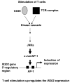
Fig. 8. Stimulation of splenic T cells activates transcription of the Ifi202 gene through the JNK/c-Jun signaling pathway

Similar articles

Cited by

References

    1. Kotzin BL. Systemic lupus erythematosus. Cell. 1996;85:303–06. - PubMed
    1. Tsokos GC, Kammer GM. Molecular aberrations in human systemic lupus erythematosus. Mol Med Today. 2000;6:418–24. - PubMed
    1. Jorgensen TN, Gubbels MR, Kotzin BL. New insights into disease pathogenesis from mouse lupus genetics. Curr Opin Immunol. 2004;16:787–93. - PubMed
    1. Theofilopoulos AN, Baccala R, Beutler B, Kono DH. Type I interferons (α/β) in immunity and autoimmunity. Annu Rev Immunol. 2005;23:307–36. - PubMed
    1. Rider V, Abdou NI. Gender differences in autoimmunity: molecular basis for estrogen effects in systemic lupus erythematosus. Int Immunopharmacol. 2001;1:1009–24. - PubMed

Publication types

MeSH terms