Review
. 2008 Jun 27;363(1500):2071-8.
doi: 10.1098/rstb.2008.2260.

Brain regeneration from pluripotent stem cells in planarian

Affiliations
Review

Brain regeneration from pluripotent stem cells in planarian

Kiyokazu Agata et al. Philos Trans R Soc Lond B Biol Sci. .

Abstract

How can planarians regenerate their brain? Recently we have identified many genes critical for this process. Brain regeneration can be divided into five steps: (1) anterior blastema formation, (2) brain rudiment formation, (3) pattern formation, (4) neural network formation, and (5) functional recovery. Here we will describe the structure and process of regeneration of the planarian brain in the first part, and then introduce genes involved in brain regeneration in the second part. Especially, we will speculate about molecular events during the early steps of brain regeneration in this review. The finding providing the greatest insight thus far is the discovery of the nou-darake (ndk; 'brains everywhere' in Japanese) gene, since brain neurons are formed throughout the entire body as a result of loss of function of the ndk gene. This finding provides a clue for elucidating the molecular and cellular mechanisms underlying brain regeneration. Here we describe the molecular action of the nou-darake gene and propose a new model to explain brain regeneration and restriction in the head region of the planarians.


Figures

Figure 1
Figure 1
Structure of the planarian CNS. (a) A whole mount in situ hybridization view from the ventral side showing the gross structure of the planarian CNS (stained with a prohormone convertase 2 homologue, DjPC2, probe). The cell bodies of neurons are stained. The planarian CNS is composed of an inverted U-shaped brain (bracket) and a pair of VNCs (arrows). (b) Immunostaining of the planarian nervous system with anti-DjSYT (planarian synaptotagmin). In contrast to the pattern in (a), only axons are stained. The ladder-like structure of the commissure neurons of the VNCs can be clearly observed (ventral view).
Figure 2
Figure 2
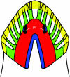
Domain structure of the planarian brain. The planarian brain can be divided into at least four structurally and functionally different domains, which are defined by the discrete expression of three otd/Otx family genes. The planarian brain is composed of two main lobes with lateral branches (nine asterisks). Lateral branch neurons are defined by Djotp expression (green) and are composed of chemosensory neurons. Both photosensory neurons and their target region in the brain are defined by DjotxA expression (blue). The two main lobes, where a variety of interneurons have been identified, are defined by DjotxB expression (red). Mechanosensory neurons (yellow) are formed in the peripheral region of the head, which is defined by being devoid of the Djotx expression of the three otd/Otx-related genes. In summary, a variety of external signals are sensed by the sensory neuron clusters in the head region, and the signals are integrated in the DjotxB-positive main lobes and transmitted to various regions of the body after processing via the pair of VNCs that are connected to the ventral side of the brain. pc, Pigmented eyecups.
Figure 3
Figure 3
Summary of brain regeneration process. Planarian brain regeneration can be divided into five steps, as indicated by the histological observations and gene expression patterns: (1) anterior blastema formation, (2) brain rudiment formation, (3) pattern formation, (4) neural network formation, and (5) functional recovery. The following genes and signalling systems are involved in these steps: step 1, BMP/noggin signal; step 2, nou-darake/FGFR signal; step 3, Wnt signal and otd/Otx family genes; step 4, netrins and CAMs; and step 5, 1020HH and eye 53, which were identified as genes involved in this step by RNAi (Inoue et al. 2004).
Figure 4
Figure 4
Brains are ectopically formed in all regions of the body in nou-darake RNAi planarians. Pluripotent stem cells distributed throughout the body give rise to brains in nou-darake RNAi planarians. The nou-darake gene encodes an FGFR-like molecule lacking a tyrosine kinase domain in its intracellular region and functions to restrict brain formation in the head region. (a) A control animal stained with a brain-specific glutamate receptor gene probe (1008HH). (bd) The nou-darake RNAi planarians stained with the same probe forming ectopic eyes and brains in the posterior region of the bodies.
Figure 5
Figure 5
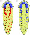
Inhibitor model. (a) The regenerating brain may secrete an inhibitor molecule that suppresses the differentiation of brain neurons in the trunk region. The brain inhibitor is indicated in red. (b) Loss of the brain inhibitor would allow the ectopic differentiation of brain neurons in the trunk region.
Figure 6
Figure 6
Interpretation of nou-darake function and its RNAi phenotype. (a) Capture model proposed in this review: a brain activator (which has not yet been identified) may stimulate the differentiation of brain neurons, but may be captured by NOU-DARAKE (NDK) and consequently not able to diffuse outside the head region. The following three experiments support the capture model: Xbra expression and gastrulation were inhibited in the planarian nou-darake mRNA-injected embryos; induction of Xbra expression in the animal cap by bFGF administration was completely suppressed in planarian nou-darake mRNA-injected animal caps; ectopic brain formation of nou-darake RNAi planarians was suppressed by co-injection of double-stranded RNAs of two FGFR homologue genes, DjFGFR1/2, which were specifically expressed in X-ray-sensitive stem cells. Brain activator and NDK are indicated in green and red, respectively. (b) In intact planarians, excess brain activators are trapped by NDK, but they can diffuse to the posterior region of the body in nou-darake RNAi planarians. (c) In triple-knockdown planarians co-injected with the two FGFR1/2 double-stranded RNAs in addition to nou-darake RNAs, ectopic brains were not formed in the trunk region due to the lack of expression of FGFR in the stem cells. The weak points of this model are that we need to postulate the existence of a third FGFR molecule (DjFGFR3), and that we have not yet identified any brain activator molecules (see text).

References

    1. Agata K. Regeneration and gene regulation in planarians. Curr. Opin. Genet. Dev. 2003;13:492–496. doi:10.1016/j.gde.2003.08.009 - DOI - PubMed
    1. Agata K, Watanabe K. Molecular and cellular aspects of planarian regeneration. Semin. Cell Dev. Biol. 1999;10:377–383. doi:10.1006/scdb.1999.0324 - DOI - PubMed
    1. Agata K, Soejima Y, Kato K, Kobayashi C, Umesono Y, Watanabe K. Structure of the planarian central nervous system (CNS) revealed by neuronal cell markers. Zool. Sci. 1998;15:433–440. doi:10.2108/zsj.15.433 - DOI - PubMed
    1. Agata K, Tanaka T, Kobayashi C, Kato K, Saitoh Y. Intercalary regeneration in planarians. Dev. Dyn. 2003;226:308–316. doi:10.1002/dvdy.10249 - DOI - PubMed
    1. Agata K, Nakajima E, Funayama N, Shibata N, Saito Y, Umesono Y. Two different evolutionary origins of stem cell systems and their molecular basis. Semin. Cell Dev. Biol. 2006;17:503–509. doi:10.1016/j.semcdb.2006.05.004 - DOI - PubMed

Publication types

MeSH terms

LinkOut - more resources