. 2008 Jun 6;283(23):15647-55.
doi: 10.1074/jbc.M800543200. Epub 2008 Mar 27.

Carcinoembryonic antigen and CD44 variant isoforms cooperate to mediate colon carcinoma cell adhesion to E- and L-selectin in shear flow

Affiliations

Carcinoembryonic antigen and CD44 variant isoforms cooperate to mediate colon carcinoma cell adhesion to E- and L-selectin in shear flow

Susan N Thomas et al. J Biol Chem. .

Abstract

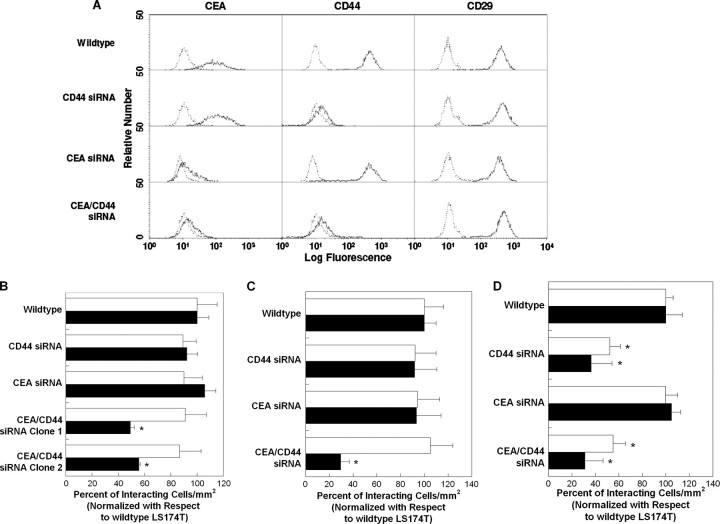
Selectin-mediated adhesion of tumor cells to platelets, leukocytes, and endothelial cells may regulate their hematogenous dissemination in the microvasculature. We recently identified CD44 variant isoforms (CD44v) as functional P-, but not E- or L-, selectin ligands on colon carcinoma cells. Moreover, an approximately 180-kDa sialofucosylated glycoprotein(s) mediated selectin binding in CD44-knockdown cells. Using immunoaffinity chromatography and tandem mass spectrometry, we identify this glycoprotein as the carcinoembryonic antigen (CEA). Blot rolling assays and flow-based adhesion assays using microbeads coated with CEA immunopurified from LS174T colon carcinoma cells and selectins as substrate reveal that CEA possesses E- and L-, but not P-, selectin ligand activity. CEA on CD44-knockdown LS174T cells exhibits higher HECA-452 immunoreactivity than CEA on wild-type cells, suggesting that CEA functions as an alternative acceptor for selectin-binding glycans. The enhanced expression of HECA-452 reactive epitopes on CEA from CD44-knockdown cells correlates with the increased CEA avidity for E- but not L-selectin. Through the generation of stable knockdown cell lines, we demonstrate that CEA serves as an auxiliary L-selectin ligand, which stabilizes L-selectin-dependent cell rolling against fluid shear. Moreover, CEA and CD44v cooperate to mediate colon carcinoma cell adhesion to E- and L-selectin at elevated shear stresses. The novel finding that CEA is an E- and L-selectin ligand may explain the enhanced metastatic potential associated with tumor cell CEA overexpression and the supportive role of selectins in metastasis.


Figures

FIGURE 1.
FIGURE 1.
Schematic diagram of the process of HECA-452 immunoaffinity chromatography, in-gel glycoprotein staining of eluted samples, tandem mass spectrometry, and bioinformatics analysis of trypsin-digested samples used to identify carcinoembryonic antigen as the 180-kDa sialofucosylated glycoprotein selectin ligand band in whole cell lysates of CD44-knockdown LS174T cells. Panel 1, HECA-452-reactive molecules were purified from the whole cell lysates of CD44-knockdown LS174T cells by immunoaffinity chromatography using KappaLock™-agarose supports cross-linked with bis(sulfosuccinimidyl)suberate to the HECA-452 mAb. HECA-452-reactive molecules were eluted using a low pH elution buffer. Panel 2, samples were then separated by SDS-PAGE and stained in-gel using ProQ Emerald 300 glycoprotein stain. The fluorescently labeled band at 180 kDa was subsequently excised from the gel. Panel 3, proteins were extracted and trypsin-digested and then subjected to tandem mass spectrometry. Bioinformatics analysis of the mass spectrometry data revealed the presence of CEA in the sample.
FIGURE 2.
FIGURE 2.
A, Western blots of whole cell lysate or immunopurified HECA-452-reactive epitopes or immunopurified CEA from CD44-knockdown LS174T colon carcinoma cells. Anti-CD66de (Col-1) (lanes 1, 3, and 5) or HECA-452 (lanes 2,4, and 6) mAbs were used to stain Western blots of CD44-knockdown LS174T whole cell lysate (lanes 1 and 2), immunoprecipitated HECA-452-reactive epitopes (lanes 3 and 4), and immunoprecipitated (IP) CEA (lanes 5 and 6) from CD44-knockdown LS174T cells. B, selectin-dependent adhesion to SDS-PAGE resolved and blotted CEA immunoprecipitated from CD44-knockdown LS174T whole cell lysate. CHO-E cells, lymphocytes, or CHO-P cells were perfused at the wall shear stress level of 0.5 dynes/cm2 over SDS-PAGE immunoblots of immunopurified CEA from whole cell lysates of CD44-knockdown LS174T cells. In select experiments, CHO-E cells and lymphocytes were pretreated with an anti-E-selectin or an anti-L-selectin function-blocking mAb (20 μg/ml), respectively, before use in blot rolling assays. The saturating concentration of the mAb (20 μg/ml) was maintained in the perfusion assays. Data represent the mean ± S.E. of n ≥ 3 experiments. ND, not done.
FIGURE 3.
FIGURE 3.
A, representative flow cytometric histograms of CEACAM expression by wild-type LS174T cells. CEACAM expression on colon carcinoma cells was investigated by using primary anti-CD66 mAbs (CD66a, GM8G5; CD66b, 80H3; CD66c, 9A6; CD66de, Col-1; CD66f, BAP3; and CEACAM7, BAC2) in conjunction with appropriate PE-conjugated secondary and isotype control antibodies. B, Western blots of whole cell lysate or immunoprecipitated (IP) CEA from wild-type LS174T colon carcinoma cells. Anti-CD66de (Col-1) (lanes 1 and 3) or HECA-452 (lanes 2 and 4) mAbs were used to stain Western blots of whole cell lysate (lanes 1 and 2) and immunoprecipitated CEA (lanes 3 and 4) from wild-type LS174T colon carcinoma cells. C, Western blots of whole cell lysate or immunoprecipitated CD66c from wild-type LS174T colon carcinoma cells. Anti-CD66c (B6.2) (lanes 1 and 3) or HECA-452 (lanes 2 and 4) mAbs were used to stain Western blots of whole cell lysate (lanes 1 and 2) and immunoprecipitated CD66c (lanes 3 and 4) from wild-type LS174T cells.
FIGURE 4.
FIGURE 4.
Site densities of CEA (A) and HECA-452-reactive epitopes (B) on polystyrene microspheres coated with CEA immunopurified from either wild-type (bold line) or CD44-knockdown (thin line) LS174T cells. Microspheres were stained with PE-conjugated anti-CD66 B1.1 (A), fluorescein isothiocyanate-conjugated HECA-452 (B), or PE- or fluorescein isothiocyanate-conjugated isotype control antibodies (dashed lines). C, extent of adhesion of microspheres (106/ml) coated with CEA immunopurified from wild-type (black bars) or CD44-knockdown (white bars) LS174T colon carcinoma cells to 10 μg/ml E-, L-, or P-selectin at a wall shear stress level of 1 dyne/cm2 for 2 min. Data represent the mean ± S.E. *, p < 0.05 with respect to microspheres coated with CEA immunopurified from wild-type LS174T cells. ND, not done. Average rolling velocities of microspheres (106/ml) coated with CEA immunopurified from wild-type or CD44-knockdown LS174T cells on 10 μg/ml E-selectin (D) or L-selectin (E) at prescribed wall shear stresses. Data represent the mean ± S.E. *, p < 0.05 with respect to microspheres coated with CEA immunopurified from wild-type LS174T cells.
FIGURE 5.
FIGURE 5.
A, representative flow cytometric histograms of CEA, CD44, and CD29 expression by wild-type, CD44-knockdown, CEA-knockdown, and CEA/CD44-double knockdown LS174T cells. Cells were stained by indirect single color immunofluorescence using the anti-CD66de mAb Col-1 (solid line) or an isotype control antibody (dashed line). Alternatively, cells were stained with the PE-conjugated anti-CD44 mAb 515 (solid line) or PE-conjugated isotype control antibody (dashed line). In other experiments, cells were stained with the PE-conjugated anti-CD29 mAb MAR4 (solid line) or PE-conjugated isotype control antibody (dashed line). B, extent of adhesion of wild-type, CD44-knockdown, CEA-knockdown, and two distinct CEA/CD44-double knockdown LS174T cell lines (106/ml) to E-selectin (0.75 μg/ml) under physiological flow conditions. The average number of wild-type LS174T cells per mm2 that tethered and rolled on E-selectin at 1.0 and 2.0 dynes/cm2 was 380 ± 60 and 290 ± 30, respectively. Data represent the mean ± S.E. of n = 3 experiments. Black bars represent data acquired at the wall shear stress level of 1.0 dyne/cm2, whereas white bars represent data at 2.0 dynes/cm2. *, p < 0.05 with respect to wild-type, CD44-knockdown, and CEA-knockdown LS174T cells. C, extent of adhesion of wild-type, CD44-knockdown, CEA-knockdown, and CEA/CD44-double knockdown LS174T cells (106/ml) to L-selectin (1.5 μg/ml) under physiological flow conditions. The average number of wild-type LS174T cells per mm2 that tethered and rolled on L-selectin at 1.0 and 2.0 dynes/cm2 was 700 ± 100 and 600 ± 100, respectively. Data are normalized with respect to wild-type LS174T cells and represent the mean ± S.E. of n = 3–4 experiments. Black bars represent data acquired at the wall shear stress level of 1.0 dyne/cm2, and white bars represent data at 2.0 dynes/cm2. *, p < 0.05 with respect to wild-type, CD44-knockdown, and CEA-knockdown LS174T cells. D, extent of adhesion of wild-type, CD44-knockdown, CEA-knockdown, and CEA/CD44-double knockdown LS174T cells (106/ml) to P-selectin (1.5μg/ml) under physiological flow conditions. The average number of wild-type LS174T cells per mm2 that tethered and rolled on P-selectin at 1.0 and 2.0 dynes/cm2 was 1200 ± 200 and 610 ± 90, respectively. Data are normalized with respect to wild-type LS174T cells and represent the mean ± S.E. of n = 3 experiments. Black bars represent data acquired at the wall shear stress level of 1.0 dynes/cm2, and white bars represent data at 2.0 dynes/cm2. *, p < 0.05 with respect to wild-type and CEA-knockdown LS174T cells.

References

    1. Hanley, W. D., Burdick, M. M., Konstantopoulos, K., and Sackstein, R. (2005) Cancer Res. 65 5812–5817 - PubMed
    1. Hanley, W. D., Napier, S. L., Burdick, M. M., Schnaar, R. L., Sackstein, R., and Konstantopoulos, K. (2006) FASEB J. 20 337–339 - PubMed
    1. Kaytes, P. S., and Geng, J. G. (1998) Biochemistry 37 10514–10521 - PubMed
    1. Kim, Y. J., Borsig, L., Han, H. L., Varki, N. M., and Varki, A. (1999) Am. J. Pathol. 155 461–472 - PMC - PubMed
    1. Mannori, G., Crottet, P., Cecconi, O., Hanasaki, K., Aruffo, A., Nelson, R. M., Varki, A., and Bevilacqua, M. P. (1995) Cancer Res. 55 4425–4431 - PubMed

Publication types

MeSH terms