. 2008 Jul;95(1):472-82.
doi: 10.1529/biophysj.107.127951. Epub 2008 Mar 28.

Insights into the mechanism of ADP action on flagellar motility derived from studies on bull sperm

Affiliations

Insights into the mechanism of ADP action on flagellar motility derived from studies on bull sperm

Kathleen A Lesich et al. Biophys J. 2008 Jul.

Abstract

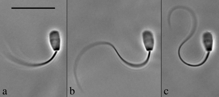
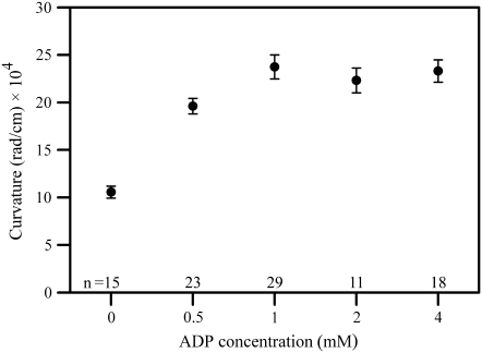
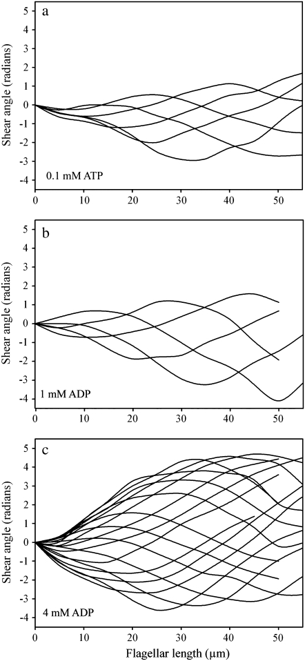
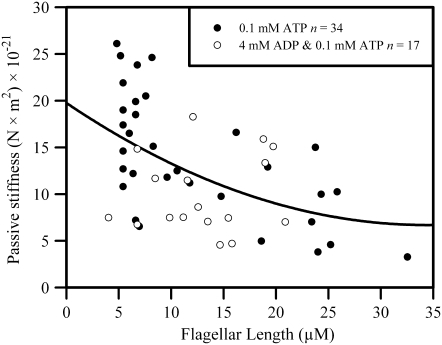
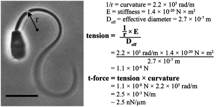
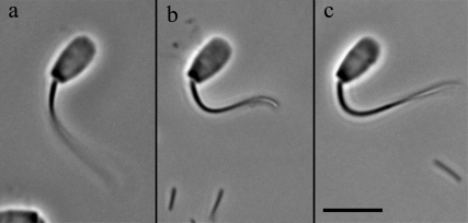
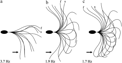
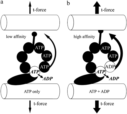
Adenosine diphosphate (ADP) is known to have interesting effects on flagellar motility. Permeabilized and reactivated bull sperm exhibit a marked reduction in beating frequency and a greatly increased beat amplitude in the presence of 1-4 mM ADP. In this study we examined the force production of sperm reactivated with 0.1 mM ATP with and without 1 mM ADP and found that there is little or no resulting change in the stalling force produced by a bull sperm flagella in response to ADP. Because bull sperm bend to a higher curvature after ADP treatment we explored the possibility that ADP-treated sperm flagella are more flexible. We measured the stiffness of 50 muM sodium vanadate treated bull sperm in the presence of 4 mM ADP, but found no change in the passive flagellar stiffness. When we analyzed the torque that develops in ADP-treated sperm at the point of beat reversal we found that the torque developed by the flagellum is significantly increased. Our torque estimates also allow us to calculate the transverse force (t-force) acting on the flagellum at the point of beat direction reversal. We find that the t-force at the switch-point of the beat is increased significantly in the ADP treated condition, averaging 0.7 +/- 0.29 nN/microm in 0.1 mM ATP and increasing to 2.9 +/- 1.2 nN/microm in 0.1 mM ATP plus 4 mM ADP. This suggests that ADP is exerting its effect on the beat by increasing the tenacity of dynein attachment at the B-subtubule. This could be a direct result of a regulatory effect of ADP on the binding affinity of dynein for the B-subtubule of the outer doublets. This result could also help to explain a number of previous experimental observations, as discussed.


Figures

FIGURE 1
FIGURE 1
The effect of ADP on the principal bend at the switch-point of the beat in bull sperm. All three images show a single frame that corresponds to the time point of greatest curvature development of the principal bend in Triton X-100 extracted cells reactivated to motility with 0.1 mM ATP. The ADP concentration is 0 mM in a, 1 mM in b, and 4 mM in c. All three cells are securely stuck to the slide surface with the flagellum beating freely. In every case, the next frame from the one shown had a reduced principal bend curvature in the region 5–10 μm from the head-tail junction and a portion of the flagellum was reversing the direction of the beat. This indicates that the selected frame is close to the switch-point of beat reversal. Bar = 20 μm.
FIGURE 2
FIGURE 2
The effect of ADP on curvature development in reactivated bull sperm. The mean (±SE) maximum curvature development of bull sperm flagella is plotted as a function of ADP concentration. Curvatures were measured between 5–10 μm from the head-tail junction on images where the flagellum was exhibiting the greatest curvature development of the principal bend in 0.1 mM ATP-reactivated sperm cells. The next frame showed a portion of the flagellum reversing the direction of the beat. As ADP content is increased from 0 to 1 mM, a significant increase in maximum curvature generation occurs (p < 0.0001; t-test). At all concentrations >1 mM ADP there is significant increased maximum curvature development (p < 0.0001) compared to cells without ADP present. There is no benefit to maximum curvature development using ADP concentrations >1 mM.
FIGURE 3
FIGURE 3
Shear angle plots for reactivated bull sperm as ADP content is varied. Each of the three plots presents the shear angle data for approximately one full beat cycle with every fourth frame plotted. All are produced from images of Triton X-100 extracted bull sperm reactivated with 0.1 mM ATP. The ADP concentration is 0 mM in a, 1 mM in b, and 4 mM in c. All of the cells were in the head stuck configuration. Note that the shear angle excursion over the course of the beat becomes progressively larger as ADP concentration increases, especially within the first 10 μm of a beating flagellum. Shear angle is the local tangent angle at a point along the flagellum minus the angle at the flagellar base. Shear angle measurements are proportional to the amount of interdoublet sliding within the axoneme.
FIGURE 4
FIGURE 4
Passive flagellar stiffness of bull sperm flagella with and without ADP. A scatter plot is shown of the accumulated determinations of flagellar stiffness on detergent extracted bull sperm that were rendered passive by 50 μM NaVO3 in the presence of 0.1 mM ATP. The flagella were bent with a force-calibrated glass microprobe. The resistance to bending was found from the force that the flagellum exerts against the calibrated microprobe when bent at various positions along the flagellar length. The stiffness, found by dividing the measured bending torque by the curvature of the induced bend, is plotted against the position of the imposed bend along the flagellum. The open circles are from measurements where 4 mM ADP was included in the solution.
FIGURE 5
FIGURE 5
Calculation of the t-force from the principal bend image of a bull sperm at the switch-point of the beat. The method we used to analyze the magnitude of the t-force, with and without ADP, is illustrated for one of the cells included in the data set. The sperm cell is reactivated with 0.1 mM ATP and is stuck to the slide by its head with the flagellum beating freely. The selected frame is the one that exhibits the greatest curvature in the region 5–10 μm from the head-tail junction (i.e., the next captured image showed less curvature and a switch in direction of beating in that same segment). The curvature is measured and the radius of curvature, r, is recorded. The radius of curvature, stiffness, and effective diameter are used in the formula shown to find the tension on the outermost axonemal component that is inducing the bend (see text for details). The tension on the axoneme multiplied by the curvature of the bend yields the global component of the t-force acting across the axoneme. The actual calculation for the specific cell shown is given as an example. This cell was treated with 4 mM ADP and yielded a t-force of 2.5 nN/μm.
FIGURE 6
FIGURE 6
The beat switch-point in bull sperm flagella shortened by microdissection. The cells shown were reactivated with 0.1 mM ATP and shortened by microdissection with a glass microprobe. The frames displayed are the images exhibiting the greatest curvature in the beat. The reactivation medium included 0 mM ADP (a), 1 mM ADP (b), and 4 mM ADP (c), respectively. Note that the curvature of the most strongly bent part of the flagellum is similar to the principal bends of the intact cells shown in Fig. 1. The same analysis as illustrated in Fig. 5 was used to estimate the t-force on cut flagella and the results are summarized in Table 3. The shortened sperm have only one bend present along the entire length of flagellum. Because of this, the entire flagellum is transiently stationary at the end point of principal bend formation and consequently has zero velocity relative to the surrounding fluid. The data from the clipped sperm show that the assumption of zero viscous drag in the analysis of t-force on the intact cells was justified and gives an additional validation of the effect of ADP on the t-force. Bar = 10 μm.
FIGURE 7
FIGURE 7
The effect of varying dynein adhesion in the Geometric Clutch computer model. The version of the Geometric Clutch model specifically developed to simulate a bull sperm flagellum (24) was used to explore the effect of dynein adhesion on the simulated beat. Three panels show the result of executing the model program, in the head stuck configuration, while varying only the scaling factor for dynein adhesion. The scaling factor was increased by 15% between a and b and by an additional 5% between b and c. The scaling factor determines what fraction of the dynein force in the model is used to reduce the probability of detachment of the dynein bridges. An increase of this single parameter of the model, while holding all other modeling parameters constant, produces some of the same changes that are actually seen in reactivated bull sperm when the ADP concentration is increased. There is a strong increase in the curvature of the principal bend at the switch-point frame (arrows). There is also an increase in the amplitude of the beat and a similar reduction in the beat frequency, as exhibited by the real sperm. In the context of the Geometric Clutch mechanism, this indicates that ADP may be exerting its action on the beat cycle by making the dynein motors more difficult to detach from their binding sites on the adjacent doublet.
FIGURE 8
FIGURE 8
A hypothetical mechanism relating ADP, dynein, and t-force. This illustration is based on the proposal put forth by Inoue and Shingyoji (17), and supported by our results presented in this study. The binding of ADP to a long residency regulatory site on the globular domain of the dynein head increases the force transfer to the neighboring doublet by enhancing the affinity of tubulin binding at the tip of the dynein stalk. This may be accomplished by enhanced transfer of a coordinating signal through the multiple p-loops of the AAA domain and/or an enhanced stability of the complete 3-D structure. The ultimate consequence, as relates to our work, is that the t-force that can be held and transferred through the stalk-tubule binding site is also affected, and therefore the release point to switch the dynein “off” is greatly increased. This results in the modification of the beat cycle to a higher t-force threshold for switching, consistent with what we see experimentally.

Similar articles

Cited by

References

    1. Lindemann, C. B., and R. Rikmenspoel. 1972. Sperm flagellar motion maintained by ADP. Exp. Cell Res. 73:255–259. - PubMed
    1. Lindemann, C. B., and I. R. Gibbons. 1975. Adenosine triphosphate-induced motility and sliding of filaments in mammalian sperm extracted with Triton X-100. J. Cell Biol. 65:147–162. - PMC - PubMed
    1. Okuno, M., and C. J. Brokaw. 1979. Inhibition of movement of trition-demembranated sea-urchin sperm flagella by Mg2+, ATP4−, ADP and Pi. J. Cell Sci. 38:105–123. - PubMed
    1. Omoto, C. K., T. Yagi, E. Kurimoto, and R. Kamiya. 1996. Ability of paralyzed flagella mutants of Chlamydomonas to move. Cell Motil. Cytoskeleton. 33:88–94. - PubMed
    1. Frey, E., C. J. Brokaw, and C. K. Omoto. 1997. Reactivation at low ATP distinguishes among classes of paralyzed flagella mutants. Cell Motil. Cytoskeleton. 38:91–99. - PubMed

Substances

LinkOut - more resources