. 2008 Jul;95(2):753-60.
doi: 10.1529/biophysj.107.125294. Epub 2008 Mar 28.

A long-lived M-like state of phoborhodopsin that mimics the active state

Affiliations

A long-lived M-like state of phoborhodopsin that mimics the active state

Yuki Sudo et al. Biophys J. 2008 Jul.

Abstract

Pharaonis phoborhodopsin (ppR, also called pharaonis sensory rhodopsin II) is a seven transmembrane helical retinal protein. ppR forms a signaling complex with pharaonis Halobacterial transducer II (pHtrII) in the membrane that transmits a light signal to the sensory system in the cytoplasm. The M-state during the photocycle of ppR (lambda(max) = 386 nm) is one of the active (signaling) intermediates. However, progress in characterizing the M-state at physiological temperature has been slow because its lifetime is very short (decay half-time is approximately 1 s). In this study, we identify a highly stable photoproduct that can be trapped at room temperature in buffer solution containing n-octyl-beta-d-glucoside, with a decay half-time and an absorption maximum of approximately 2 h and 386 nm, respectively. HPLC analysis revealed that this stable photoproduct contains 13-cis-retinal as a chromophore. Previously, we reported that water-soluble hydroxylamine reacts selectively with the M-state, and we found that this stable photoproduct also reacts selectively with that reagent. These results suggest that the physical properties of the stable photoproduct (named the M-like state) are very similar with the M-state during the photocycle. By utilizing the high stability of the M-like state, we analyzed interactions of the M-like state and directly estimated the pK(a) value of the Schiff base in the M-like state. These results suggest that the dissociation constant of the ppR(M-like)/pHtrII complex greatly increases (to 5 muM) as the pK(a) value greatly decreases (from 12 to 1.5). The proton transfer reaction of ppR from the cytoplasmic to the extracellular side is proposed to be caused by this change in pK(a).


Figures

FIGURE 1
FIGURE 1
Light-induced spectral changes of ppR. (a) Time-dependent spectral changes of ppR. The spectra were recorded at 0, 20, 50, 80, and 120 min. Spectra except for 0 min were noisy because we monitored these spectra under constant light illumination. (b) Difference absorption spectra. The reaction mixture was irradiated with an intensity of 5 W/m2. ppR at 18 μM was suspended in buffer at pH 5.0 (see Materials and Methods) at 25°C.
FIGURE 2
FIGURE 2
Spectral transition from a photoproduct (λmax = 386 nm) to the ground state (λmax = 500 nm) of ppR. (a) Time-dependent spectral changes of ppR. The spectra were recorded at 0, 60, 120, 540, and 1200 min. (b) Difference absorption spectra. Experimental conditions were as described for Fig. 1.
FIGURE 3
FIGURE 3
Chromophore configuration of the photoproduct (the M-like state). The detection beam was set at 360 nm. Samples were collected and extracted from the protein at 0, 20, 60, 80, and 120 min. The fraction of 13-cis- and all-trans-retinal was determined by calculation of molar compositions from the areas of the peaks in the HPLC patterns. Solid lines that indicate light-induced absorbance changes were reproduced from Fig. 3 a. The fraction of 13-cis- and all-trans-retinal were well correlated with absorbance changes of the decrease of the ground state and the increase of the photoproduct (the M-like state).
FIGURE 4
FIGURE 4
(a) Photoinduced difference spectra of ppRM-like. (b) Reactivity of the ground states of ppR (open circles) and ppRM-like (solid circles) with hydroxylamine. For ppRM-like, 50 mM hydroxylamine was added in buffer solution after constant illumination of ppR for 120 min. For the ground state of ppR, 50 mM hydroxylamine was also added in buffer solution. For measurement of the reaction of ppR with hydroxylamine, time-dependent absorbance changes at 345 nm were monitored. The slopes are: 0.0026 min−1 for the M-like state and 0.0004 min−1 for the ground state.
FIGURE 5
FIGURE 5
(a) Spectral transition from the M-like state to the ground state in the presence (solid circles) or absence of pHtrII (open circles). ppR samples (8 μM) with or without pHtrII (40 μM) were irradiated by yellow light through a Y52 filter for 360 min. The temperature was kept at 25°C. The reverse reaction from the M-like state to the ground state in the presence or absence of purified pHtrII was monitored using a UV-Vis spectrometer as an increase in absorbance at 500 nm. (b) Interaction between ppRM-like and ppR. The concentration of free ppRM-lilke, [ppRM-like], and the ppRM-lilke/pHtrII complex, [ppRM-lilke/pHtrII], were calculated according to Eqs. 1–3 and are expressed as micromolar. The solid line represents the fit curve. Data were regressed with nonlinear regression Origin software to evaluate KD and n, which were 5.2 μM and 0.7, respectively.
FIGURE 6
FIGURE 6
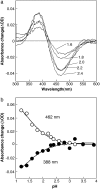
Estimation of pKa value of the Schiff base. (a) pH titration of the Schiff base in ppRM-like. Changes in absorption spectra of protonated M-like state from 1.2 to 3.9 were monitored. The pH values of each spectrum are 2.4, 2.2, 2.0, 1.8, and 1.6, respectively. (a and b) Estimated absorbance changes using spectral deconvolution at 388 and 462 nm, respectively. Experimental conditions were as reported for Fig. 1 except for the salt concentration (0 mM). The plots are based on three or four independent experiments.

References

    1. Spudich, J. L., C.-S. Yang, K.-H. Jung, and E. N. Spudich. 2000. Retinylidene proteins: structures and functions from archaea to humans. Annu. Rev. Cell Biol. 16:365–392. - PubMed
    1. Sudo, Y., H. Kandori, and N. Kamo. 2004. Molecular mechanism of protein-protein interaction of pharaonis phoborhodopsin/transducer and photo-signal transfer reaction by the complex. Recent Res. Devel. Biophys. 3:1–16.
    1. Falke, J. J., R. B. Bass, S. L. Butler, S. A. Chervitz, and M. A. Danielson. 1997. The two-component signaling pathway of bacterial chemotaxis: a molecular view of signal transduction by receptors, kinases, and adaptation enzymes. Annu. Rev. Cell Dev. Biol. 13:457–512. - PMC - PubMed
    1. Rudolph, J., and D. Oesterhelt. 1996. Deletion analysis of the che operon in the archaeon Halobacterium salinarium. J. Mol. Biol. 258:548–554. - PubMed
    1. Ikeura, Y., K. Shimono, M. Iwamoto, Y. Sudo, and N. Kamo. 2003. Arg-72 of pharaonis phoborhodopsin (sensory rhodopsin II) is important for the maintenance of the protein structure in the solubilized state. Photochem. Photobiol. 77:96–100. - PubMed

Publication types

MeSH terms

Substances

LinkOut - more resources