. 2008 Aug;22(8):2662-75.
doi: 10.1096/fj.07-097709. Epub 2008 Mar 28.

Transglutaminase 2 protects against ischemic insult, interacts with HIF1beta, and attenuates HIF1 signaling

Affiliations

Transglutaminase 2 protects against ischemic insult, interacts with HIF1beta, and attenuates HIF1 signaling

Anthony J Filiano et al. FASEB J. 2008 Aug.

Abstract

Transglutaminase 2 (TG2) is a multifunctional enzyme that has been implicated in the pathogenesis of neurodegenerative diseases, ischemia, and stroke. The mechanism by which TG2 modulates disease progression have not been elucidated. In this study we investigate the role of TG2 in the cellular response to ischemia and hypoxia. TG2 is up-regulated in neurons exposed to oxygen and glucose deprivation (OGD), and increased TG2 expression protects neurons against OGD-induced cell death independent of its transamidating activity. We identified hypoxia inducible factor 1beta (HIF1beta) as a TG2 binding partner. HIF1beta and HIF1alpha together form the heterodimeric transcription factor hypoxia inducible factor 1 (HIF1). TG2 and the transaminase-inactive mutant C277S-TG2 inhibited a HIF-dependent transcription reporter assay under hypoxic conditions without affecting nuclear protein levels for HIF1alpha or HIF1beta, their ability to form the HIF1 heterodimeric transcription factor, or HIF1 binding to its DNA response element. Interestingly, TG2 attenuates the up-regulation of the HIF-dependent proapoptotic gene Bnip3 in response to OGD but had no effect on the expression of VEGF, which has been linked to prosurvival processes. This study demonstrates for the first time that TG2 protects against OGD, interacts with HIF1beta, and attenuates the HIF1 hypoxic response pathway. These results indicate that TG2 may play an important role in protecting against the delayed neuronal cell death in ischemia and stroke.


Figures
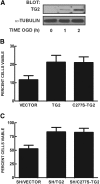
Figure 1.
Figure 1.
TG2 is up-regulated after OGD, and increased expression of TG2 is neuroprotective. A, B) Rat primary cortical neurons were treated with OGD for 1 or 2 h at 12 DIV. Cells were placed back in conditioned medium and ambient culture chambers for 24 h before collection. A) Immunoblots show that endogenous rat TG2 is up-regulated 24 h after 1 or 2 h of OGD. Blots were reprobed with α-tubulin to show equivalent loading. B) Transient transfection of TG2 and C227S-TG significantly protects rat primary neurons from 2 h of OGD (P<0.05). C) SH-SY5Y cells that stably overexpress TG2 (SH/TG2 or SH/C277S-TG2) are significantly more resistant to cell death induced by 12 h of OGD (P<0.05).
Figure 2.
Figure 2.
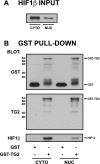
TG2 interacts with HIF1β in vitro. A GST pull-down assay was performed using recombinant GST-TG2 and HIF1β from transiently transfected CHO cell nuclear (NUC) and cytosolic (CYTO) fractions. A) Immunoblot for HIF1β protein in cytosolic and nuclear fractions. B) Cytosolic and nuclear lysates were incubated with glutathione beads that had been prebound with either GST or GST-TG2. Immunoblot analysis for GST (top panel) demonstrates that GST (lanes 1 and 3) and GST-TG2 (lanes 2 and 4) had bound to beads, and immunoblot analysis for TG2 (middle panel) confirms that GST-TG2 (lanes 2 and 4) had bound to beads in this experiment. Immunoblot analysis for HIF1β within the same samples shows that HIF1β was pulled down by GST-TG2 but not by GST alone.
Figure 3.
Figure 3.
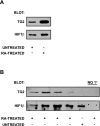
Endogenous TG2 coimmunoprecipitates with endogenous HIF1β in situ. SH-SY5Y cells were treated with 10 μM all-trans retinoic acid (RA) for 5 days and collected. A) Immunoblots of cells incubated in the absence or presence of RA for 5 days show that TG2 is up-regulated in response to RA treatment. Blots were also probed for HIF1β. B) Four samples of RA-treated cells from 2 independent experiments were immunoprecipitated for HIF1β and immunoblotted for both HIF1β and TG2. The up-regulation of TG2 in response to RA treatment resulted in a detectable interaction between TG2 and HIF1β in all the samples. The last lane shows a control immunoprecipitation in which the immunoprecipitating antibody was omitted.
Figure 4.
Figure 4.
TG2 attenuates HIF1-dependent transcriptional activity in situ. SH/vector, SH/TG2, and SH/C277S-TG2 cells were transfected with constructs for firefly luciferase (under control of the HRE promoter) and Renilla luciferase (control). Each cell type was then divided into one of four experimental conditions as indicated, based on growth conditions and the exogenous expression of HIF1β. All data are expressed as the fold increase compared with SH/vector cells grown under normoxic conditions. Luciferase activity did not differ among the 3 cell types when grown under normoxic conditions, regardless of whether HIF1β was exogenously expressed (open and checked bars). For cells grown under hypoxia but without exogenous HIF1β expression (gray bars), HRE activity was decreased in SH/C277S-TG2 cells compared with both SH/vector and SH/TG2 cells (P<0.05), whereas HRE activity did not differ between SH/vector and SH/TG2 cells (P>0.05). For cells grown under hypoxia with exogenous HIF1β expression (solid bars), HRE activity was reduced in both SH/TG2 and SH/C277S-TG2 cells compared with SH/vector cells (P<0.05). HRE activity was not statistically different between SH/TG2 and SH/C277S-TG2 cells (P>0.05).
Figure 5.
Figure 5.
TG2 differentially affects the expression of HIF-dependent transcripts. SH/vector, SH/TG2, and SH/C277S-TG2 cells were treated under the same conditions as in the cell death studies (normoxia vs. 12 h OGD) before mRNA collection and generation of cDNA. VEGF and Bnip3 genes were analyzed using Q-RT-PCR normalized to β-actin to control for total mRNA collected. All data are presented as fold induction after OGD. No significant difference in VEGF mRNA levels after OGD was observed in SH/TG2 and SH/C277S-TG2 cells when compared with SH/vector cells (P>0.05). However, a significant attenuation of Bnip3 levels in SH/TG2 and SH/C277S-TG2 was found compared to SH/vector (P<0.05).
Figure 6.
Figure 6.
TG2 attenuates HIF transcriptional activity in an HIF1α-independent manner. Human renal carcinoma 786-0 cells, which lack a functional VHL and predominantly express HIF2α over HIF1α, were transfected with constructs for firefly luciferase (under control of the HRE promoter) and Renilla luciferase (control) along with either wild-type TG2, inactive mutant C277S-TG2, or pcDNA 3.1(+) vector control. Cells were allowed to express protein for 24 h in ambient air incubators then collected and HRE activity measured. All data are expressed as the fold increase compared with vector transfected cells. HRE activity was significantly reduced by both wild TG2 and C277S-TG2 compared to vector controls (P<0.001).
Figure 7.
Figure 7.
TG2 does not attenuate HIF1α or HIF1β localization to the nucleus under hypoxia. SH/vector, SH/TG2, and SH/C277S-TG2 cells were grown under either normoxic (–) or hypoxic (+) conditions. Cells were collected, and nuclear and cytosolic fractions were separated. A) Immunoblot analysis of the nuclear fractions demonstrates that under hypoxic conditions nuclear HIF1α protein content is greater in SH/TG2 cells compared with SH/vector cells and that nuclear HIF1α protein content is increased further still in SH/C277S-TG2 cells (top panel). Although nuclear HIF1β protein content was greater for all cell types grown under hypoxic conditions compared with normoxic conditions, TG2 did not affect nuclear HIF1β protein content for cells grown in hypoxia (middle panel). TG2 protein content is also shown (bottom panel). B) Nuclear and cytosolic fractions were immunoblotted for α-tubulin as a cytosolic marker and histone subunit H4 as a nuclear marker. These immunoblots show that the nuclear fractions are not contaminated with cytosolic protein in each of the 3 cell lines.
Figure 8.
Figure 8.
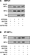
TG2 does not affect the interaction between nuclear HIF1α and HIF1β under hypoxia. SH/vector, SH/TG2, and SH/C277S-TG2 cells were grown under either normoxic or hypoxic conditions. Cells were collected, and nuclear fractions were isolated. A) Protein content for HIF1α (top panel), HIF1β (middle panel), and TG2 (bottom panel) in 15 μg nuclear protein from the indicated cell type. B) HIF1α was immunoprecipitated from 25 μg nuclear protein that was collected from the indicated cell types. Immunoprecipitated protein was immunoblotted for HIF1α (top panel) and HIF1β (bottom panel). TG2 did not affect the ability of HIF1β to coimmunoprecipitate with HIF1α. Last lane shows a sham immunoprecipitation in which the immunoprecipitating antibody was omitted.
Figure 9.
Figure 9.
TG2 does not attenuate HIF1 binding to the HRE under hypoxia. SH/vector, SH/TG2, and SH/C277S-TG2 cells were grown under either normoxic or hypoxic conditions. Cells were collected, and nuclear HIF1 binding to the HRE was measured using a modified ELISA-based assay. HIF1 binding to the HRE was increased significantly for each cell type grown in hypoxic conditions compared with normoxic conditions (P<0.0001). Although HIF1 binding to the HRE was increased in SH/TG2 cells grown under normoxia compared with SH/vector and SH/C277S-TG2 cells (P<0.001), no effect of TG2 or C277S-TG2 on HIF1 binding for cells grown under hypoxic conditions was detected (P>0.05).

Similar articles

Cited by

References

    1. Lorand L, Graham R M. Transglutaminases: crosslinking enzymes with pleiotropic functions. Nat Rev Mol Cell Biol. 2003;4:140–156. - PubMed
    1. Achyuthan K E, Greenberg C S. Identification of a guanosine triphosphate-binding site on guinea pig liver transglutaminase. Role of GTP and calcium ions in modulating activity. J Biol Chem. 1987;262:1901–1906. - PubMed
    1. Nakaoka H, Perez D M, Baek K J, Das T, Husain A, Misono K, Im M J, Graham R M. Gh: a GTP-binding protein with transglutaminase activity and receptor signaling function. Science. 1994;264:1593–1596. - PubMed
    1. Lee K N, Birckbichler P J, Patterson M K., Jr GTP hydrolysis by guinea pig liver transglutaminase. Biochem Biophys Res Commun. 1989;162:1370–1375. - PubMed
    1. Hasegawa G, Suwa M, Ichikawa Y, Ohtsuka T, Kumagai S, Kikuchi M, Sato Y, Saito Y. A novel function of tissue-type transglutaminase: protein disulphide isomerase. Biochem J. 2003;373:793–803. - PMC - PubMed

Publication types

MeSH terms

LinkOut - more resources