. 2008 Jun;190(11):3877-85.
doi: 10.1128/JB.00209-08. Epub 2008 Mar 28.

Transcriptional analysis and functional characterization of a gene pair encoding iron-regulated xenocin and immunity proteins of Xenorhabdus nematophila

Affiliations

Transcriptional analysis and functional characterization of a gene pair encoding iron-regulated xenocin and immunity proteins of Xenorhabdus nematophila

Jitendra Singh et al. J Bacteriol. 2008 Jun.

Abstract

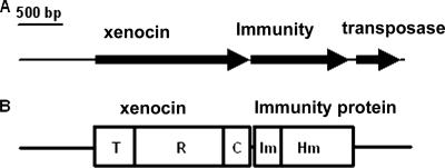
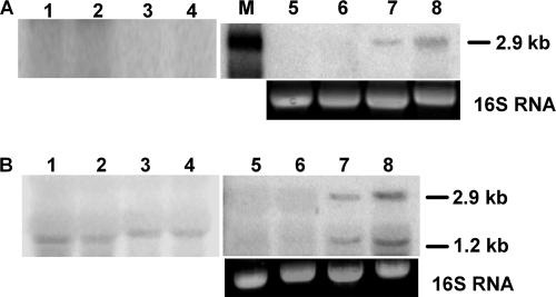
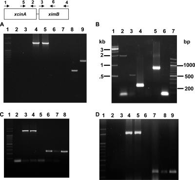
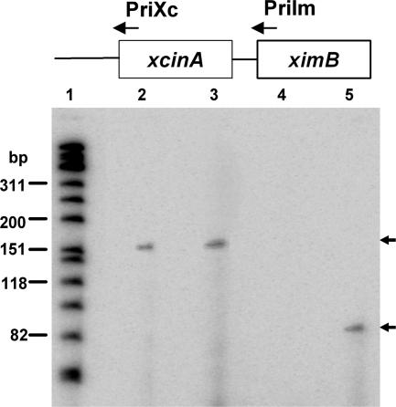
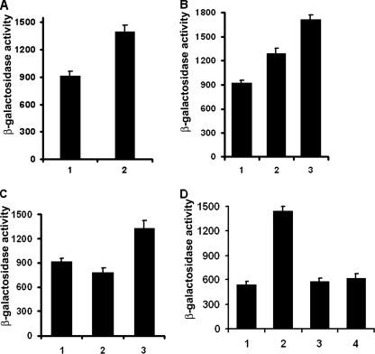
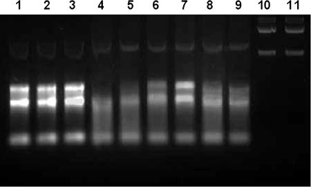
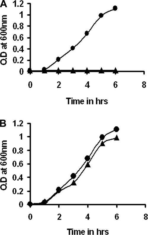
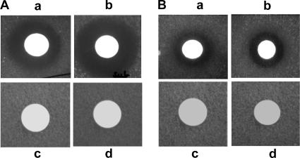
We describe a two-gene cluster encoding a bacteriocin, xenocin, and the cognate immunity protein in the insect-pathogenic bacterium Xenorhabdus nematophila, which infects and kills larval stages of the common crop pest Helicoverpa armigera. The two genes, xcinA and ximB, are present in the genome as a single transcriptional unit, which is regulated under SOS conditions. The stress-inducible promoter was activated by mitomycin C, glucose, and Fe(3+) depletion and at an elevated temperature when it was tested in Escherichia coli cells. Expression of the xenocin protein alone in E. coli inhibited the growth of this organism. The growth inhibition was abolished when the immunity protein was also present. A recombinant xenocin-immunity protein complex inhibited the growth of E. coli indicator cells when it was added exogenously to a growing culture. Xenocin is an endoribonuclease with an enzymatically active C-terminal domain. Six resident bacterial species (i.e., Bacillus, Enterobacter, Enterococcus, Citrobacter, Serratia, and Stenotrophomonas species) from the H. armigera gut exhibited sensitivity to recombinant xenocin when the organisms were grown under iron-depleted conditions and at a high temperature. Xenocin also inhibited the growth of two Xenorhabdus isolates. This study demonstrates that Fe(3+) depletion acts as a common cue for synthesis of xenocin by X. nematophila and sensitization of the target strains to the bacteriocin.


Figures

FIG. 1.
FIG. 1.
(A) Genomic organization of X. nematophila xenocin gene cluster (4,337 bp). The arrows indicate transcriptional polarities of the ORFs encoding xenocin, the immunity protein, and the transposase protein. (B) Domain map of the encoded proteins. T, translocation domain; R, receptor binding domain; C, catalytic domain; Im, immunity domain; Hm, hemolysin domain.
FIG. 2.
FIG. 2.
Northern blot of xcinA and ximB genes. Total RNA was obtained from uninduced and mitomycin C-induced X. nematophila cells at different times. Ten micrograms of RNA was used in each lane. (A) Blot probed with labeled xcinA gene. Lanes 1 to 4, RNA from uninduced cells at 0, 1, 2, and 3 h, respectively, with ethidium bromide-stained bands corresponding to the 16S RNA of the gel as a loading control; lanes 5 to 8, RNA from mitomycin C-induced cells at 0, 1, 2, and 3 h, respectively; lane M, 3-kb RNA marker. (B) Blot probed with labeled xcimB gene. Lanes 1 to 4, RNA from uninduced cells at 0, 1, 2, and 3 h, respectively; lanes 5 to 8, RNA from mitomycin C-induced cells at 0, 1, 2, and 3 h, respectively, with ethidium bromide-stained bands corresponding to 16S RNA in the gel used as a loading control.
FIG. 3.
FIG. 3.
Analysis of xcinA and ximB mRNA by RT-PCR. (A) Comparison of X. nematophila strain 19060 and xenocin-sensitive isolate X(sensitive) after induction with mitomycin C. Lane 1, markers; lanes 2 and 3, RNA from X(sensitive) and strain 19060, respectively, without reverse transcriptase; lanes 4 and 5, control 16S RNA from X(sensitive) and strain 19060, respectively; lanes 6 and 7, RNA from X(sensitive) amplified with primers 5 and 2 (lane 6) and with primers 5 and 6 (lane 7); lanes 8 and 9, RNA from X. nematophila 19060 amplified with primers 5 and 2 (lane 8) and with primers 5 and 6 (lane 9). (B) X. nematophila cells were induced with mitomycin C for 3 h, and total RNA was isolated. Lane 1, 1-kb ladder; lane 2, primers 1 and 2; lane 3, primers 5 and 6; lane 4, primers 5 and 2; lane 5, primers 3 and 4; lane 6, primers 1 and 4; lane 7, 100-bp ladder. (C) Analysis of xcinA mRNA induced by 25 mM Desferal. Lane 1, 100-bp ladder; lane 2, RNA from induced cells without reverse transcriptase, amplified with primers 5 and 2; lanes 3 and 4, 16S RNA from uninduced and induced loading controls, respectively; lanes 5 and 6, RNA from uninduced (lane 5) and induced (lane 6) cells amplified with primers 5 and 2; lanes 7 and 8, RNA from uninduced (lane 7) and induced (lane 8) cells amplified with primers 3 and 6. (D) Analysis of xcinA mRNA by heat induction. Lane 1, 100-bp ladder; lanes 2 and 3, RNA from uninduced and induced cells without reverse transcriptase, respectively; lanes 4 and 5, 16S RNA controls; lanes 6 and 7, RNA from cells grown at 30°C (lane 6) and at 37°C (lane 7) amplified with primers 5 and 2; lanes 8 and 9, RNA from cells grown at 30°C (lane 8) and at 37°C (lane 9) amplified with primers 3 and 6.
FIG. 4.
FIG. 4.
Promoter identification by primer extension. Total RNA was obtained from uninduced and mitomycin C-induced X. nematophila cells. Each reaction mixture contained 40 μg RNA. Lane 1, radiolabeled φX174 DNA marker; lanes 2 and 3, PriIm primer from the N′ terminus of the ximB gene with RNA from uninduced and induced cells, respectively; lanes 4 and 5, PriXc primer from the N′ terminus of the xcinA gene with RNA from uninduced and induced cells, respectively.
FIG. 5.
FIG. 5.
β-Galactosidase activities of xcinA promoter-lacZ fusions. E. coli strains were grown to log phase in LB medium and resuspended in M9 medium under specific nutrient conditions for induction. (A) Cells grown in M9 medium. Bar 1, no mitomycin C; bar 2, induction with 0.3 μg/ml mitomycin C. (B) Activities at different temperatures. Bar 1, 30°C; bar 2, 37°C; bar 3, 42°C. (C) Cells grown in M9 media. Bar 1, M9 medium control; bar 2, M9 medium with 2 μg/ml FeSO4; bar 3, M9 medium with 2 μg/ml FeSO4 and 25 mM Desferal. (D) Effect of glucose. Bars 1 and 2, no glucose at 0 and 2 h, respectively; bars 3 and 4, cells with 0.2% glucose at 0 and 2 h, respectively. The data are expressed in Miller units.
FIG. 6.
FIG. 6.
RNase activity of the purified catalytic domain. Each 20-μl reaction mixture containing 1.2 μg of RNA with the test protein was incubated at 37°C for 1.5 h. Lane 1, RNA without protein; lane 2, RNA with 10 μg of catalytic domain-immunity domain complex; lane 3, 10 μg heat-inactivated catalytic domain; lanes 4, 5, and 6, RNA with 10, 5, and 1 μg catalytic domain, respectively; lanes 7 and 8, 5 μg catalytic domain-immunity domain protein at molar ratios of 1:3 and 1: 1, respectively; lane 9, 5 μg catalytic domain-bovine serum albumin at a molar ratio of 1:2; lane 10, E. coli DNA without protein; lane 11, DNA with 5 μg catalytic domain protein.
FIG. 7.
FIG. 7.
Growth curves for strains BSK1 and BSK2. Cultures were grown to log phase and subcultured in fresh medium with and without mitomycin C. Bacterial growth was monitored by determining the optical density at 600 nm (O.D at 600 nm). (A) •, uninduced strain BSK2; ▴, strain BSK2 induced with mitomycin C. (B) •, uninduced strain BSK1; ▴, induced BSK1 cells.
FIG. 8.
FIG. 8.
Exogenous bacteriostatic activity. (A) Zone of clearance of BL21(DE3)/pLysS target cells grown in M9 medium with (a) cytoplasmic and (b) supernatant fractions of BSK1 cells after induction with mitomycin C. BL21(DE3)/pLysS target cells were also grown in M9 minimal medium containing iron and tested with (c) cytoplasmic and (d) supernatant fractions of mitomycin C-induced BSK4.3 cells. (B) Zone of clearance of (a) DH5α and (b) BL21(DE3)/pLysS target cells with supernatant of JC2 cells after induction with IPTG, (c) induced supernatant of M15 cells containing vector alone, and (d) buffer with BL21(DE3)/pLysS target cells.

References

    1. Akhurst, R. J. 1982. Antibiotic activity of Xenorhabdus spp. bacteria symbiotically associated with insect pathogenic nematodes of the families Heterorhabditae and Steinernematidae. J. Gen. Microbiol. 1283061-3065. - PubMed
    1. Akhurst, R. J., and G. B. Dunphy. 1993. Symbiotically associated entomopathogenic bacteria, nematodes and their insect hosts, p. 1-23. In N. Beckage, S. Thompson, and B. Federici (ed.), Parasites and pathogens of insects, vol. 2. Academic Press, Inc., New York, NY.
    1. Ausbel, F. M., R. Brent, R. E. Kingston, D. D. Moore, J. G. Seidman, J. A. Smith, and K. Struhl (ed.). 1989. Current protocols in molecular biology. John Wiley and Sons, New York, NY.
    1. Barnard, T. J., M. E. Watson, Jr., and M. A. McIntosh. 2001. Mutations in the Escherichia coli receptor FepA reveal residues involved in ligand binding and transport. Mol. Microbiol. 41527-536. - PubMed
    1. Bedding, R. A., and R. J. Akhrust. 1975. Nematodes and their biological control of insect pests. Nematologia 21109-110.

Publication types

MeSH terms

LinkOut - more resources