. 2008 Apr;26(4):453-61.
doi: 10.1038/nbt1395. Epub 2008 Mar 30.

Tumor immunotherapy across MHC barriers using allogeneic T-cell precursors

Affiliations

Tumor immunotherapy across MHC barriers using allogeneic T-cell precursors

Johannes L Zakrzewski et al. Nat Biotechnol. 2008 Apr.

Erratum in

  • Author Correction: Tumor immunotherapy across MHC barriers using allogeneic T-cell precursors.
    Zakrzewski JL, Suh D, Markley JC, Smith OM, King C, Goldberg GL, Jenq R, Holland AM, Grubin J, Cabrera-Perez J, Brentjens RJ, Lu SX, Rizzuto G, Sant'Angelo DB, Riviere I, Sadelain M, Heller G, Zúñiga-Pflücker JC, Lu C, van den Brink MRM. Zakrzewski JL, et al. Nat Biotechnol. 2024 Apr;42(4):674. doi: 10.1038/s41587-024-02205-3. Nat Biotechnol. 2024. PMID: 38531977 No abstract available.

Abstract

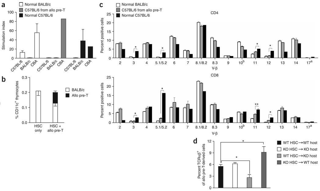
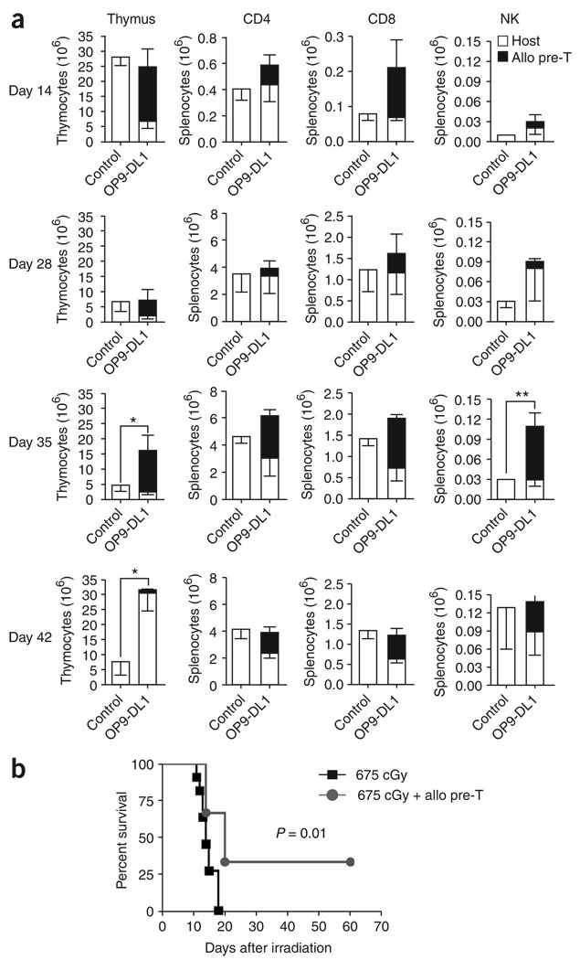
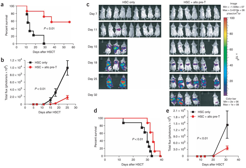
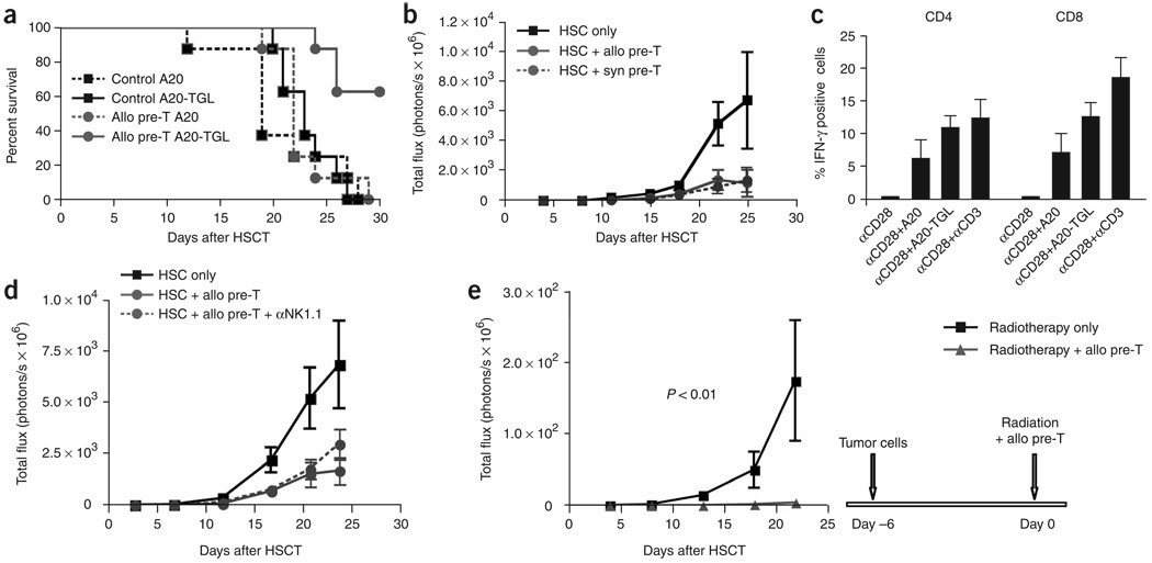
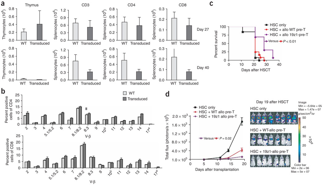
We present a strategy for adoptive immunotherapy using T-lineage committed lymphoid precursor cells generated by Notch1-based culture. We found that allogeneic T-cell precursors can be transferred to irradiated individuals irrespective of major histocompatibility complex (MHC) disparities and give rise to host-MHC restricted and host-tolerant functional allogeneic T cells, improving survival in irradiated recipients as well as enhancing anti-tumor responses. T-cell precursors transduced to express a chimeric receptor targeting hCD19 resulted in significant additional anti-tumor activity, demonstrating the feasibility of genetic engineering of these cells. We conclude that ex vivo generated MHC-disparate T-cell precursors from any donor can be used universally for 'off-the-shelf' immunotherapy, and can be further enhanced by genetic engineering for targeted immunotherapy.


Figures

Figure 1
Figure 1
Adoptively transferred allogeneic T-cell precursors enhance T and NK cell reconstitution and do not induce GVHD. (a) Lethally irradiated BALB/c mice were transplanted with syngeneic purified HS cells (or lin bone marrow cells); control mice received HS cells only, the treatment group received additional T-cell precursors generated in OP9-DL1 cocultures. In some cases, transplantation recipients were intravenously challenged with tumor cells to study anti-tumor activity. allo, allogeneic. (b) Lethally irradiated BALB/c recipients were transplanted with BALB/c mouse HS cells (Ly9.1); control mice received HS cells only, the treatment group received additional C57BL/6-derived in vitro–generated T-cell precursors (CD45.1+). At days 14, 28 and 60 after HSCT, animals were killed and thymi and spleens were harvested. Origin of cells, whether from BALB/c or C57BL/6 mice, was determined by total cellularity and multicolor flow cytometric analysis using Ly9.1- and CD45.1-specific antibodies. T and NK cells were analyzed using antibodies to CD3, CD4, and CD8, and DX5, respectively. Combined data of more than three independent experiments are presented. Values represent mean cell numbers and s.e.m. (n = 5–10). *, P < 0.05. (c) Lethally irradiated BALB/c recipients were transplanted with lin BALB/c mouse bone marrow; control mice received bone marrow only, the treatment group received additional C57BL/6 T-cell precursors. Survival was monitored daily, a clinical GVHD score was monitored weekly. Histopathological analysis of signs of subclinical GVHD in liver, small bowel and large bowel was performed 8 weeks after transplantation. Data are representative of more than three independent experiments. Mean values and s.e.m. are presented (n = 5).
Figure 2
Figure 2
Adoptively transferred allogeneic T-cell precursors develop into host MHC restricted and donor/host tolerant T cells. (a) Lethally irradiated BALB/c recipients were transplanted with BALB/c mice HS cells and received additional C57BL/6-derived T-cell precursors. Animals were killed at day 55–60 after HSCT and purified splenic T cells of BALB/c (normal BALB/c) and C57BL/6 (C57BL/6 from allo pre-T) origin were used as effector cells in mixed leukocyte reaction cultures. C57BL/6 T-cells from untreated C57BL/6 mice (normal C57BL/6) were used as additional controls. Cells were cultured for 5 d with irradiated C57BL/6 mouse, BALB/c mouse or CBA mouse splenocytes as stimulators. Combined data of three independent experiments are presented as mean ± s.e.m. (n = 8). (b) Lethally irradiated BALB/c recipients weretransplanted with BALB/c mouse HS cells and with or without additional C57CL/6-derived in vitro-generated T-cell precursors. Animals were killed on day 14 after HSCT and thymi were harvested for multicolor flow cytometric analysis of CD11c+ cells of BALB/c and C57BL/6 origin. Mean ± s.e.m. are presented (n = 5). The experiment was repeated at least three times. (c) Lethally irradiated BALB/c recipients were transplanted with BALB/c mouse HS cells and received additional C57CL/6-derived in vitro-generated T-cell precursors. Animals were killed on day 60 after HSCT and splenocytes were obtained for multicolor flow cytometric analysis of the TCR-Vβ families on CD4+ and CD8+ cells of C57BL/6 origin. BALB/c and C57BL/6 splenocytes from untreated BALB/c and C57BL/6 mice (normal BALB/c and C57BL/6) were used as controls. Mean ± s.e.m. are presented (n = 5–8). Combined data from two independent experiments are presented. *, P < 0.01; **, P < 0.05. (d) Lethally irradiated C57BL/6 wild-type recipients or C57BL/6 MHC class I and II–deficient mice were transplanted with syngeneic MHC-positive or MHC-deficient HS cells. All animals received BALB/c-derived MHC-positive T-cell precursors on day 0. Animals were killed on day 14 after HSCT and thymocytes were obtained for multicolor flow cytometric analysis of BALB/c-derived TCRαβ+ cells. Mean ± s.e.m. of combined data from two independent experiments are presented (n = 4–6). *, P < 0.05. allo pre-T, allogeneic T-cell precursors.
Figure 3
Figure 3
Adoptively transferred allogeneic T-cell precursors enhance T and NK cell reconstitution and improve survival in irradiated hosts. (a) BALB/c recipients were irradiated with a single dose of 650–687 cGy. Control mice received irradiation only; the treatment group received additional C57BL/6-derived in vitro-generated T-cell precursors. At days 14, 28, 35 and 42 after irradiation, animals were killed and thymi and spleens were harvested. BALB/c or C57BL/6 origin of cells was determined by total cellularity and multicolor flow cytometric analysis using Ly9.1- and CD45.1-specific antibodies, respectively. T and NK cells were analyzed using antibodies to CD3, CD4, and CD8, and DX5, respectively. Values represent mean cell numbers ± s.e.m. (n = 5–12). Combined data of two independent experiments are presented. *, P < 0.05; **, P < 0.01. (b) BALB/c recipients were irradiated with a single dose of 675 cGy. Control mice received irradiation only; the treatment group received additional C57BL/6-derived in vitro-generated T-cell precursors. Survival was monitored daily. One of three independent experiments is presented (n = 6–11).
Figure 4
Figure 4
Adoptively transferred allogeneic T-cell precursors mediate significant anti-tumor responses. (a–c) Lethally irradiated BALB/c recipients were transplanted with BALB/c HS cells; control mice received HS cells only, the treatment group received additional C57BL/6-derived in vitro-generated T-cell precursors on day 0. All mice received 2.5 × 10⁵ luciferase-expressing A20-TGL tumor cells intravenously on day 0. Over the course of 32 d after injection, the whole body distribution of transduced tumor cells was monitored using in vivo bioluminescence imaging. Survival is shown in a, the bioluminescent signal intensity for every group at six time points presented as mean ± s.e.m. is shown in b and pseudo-color images superimposed on conventional photographs are shown in c (n = 7–9). (d–e) Lethally irradiated BALB/c recipients were transplanted with BALB/c HS cells; control mice received HS cells only, the treatment group received additional C57BL/6-derived in vitro-generated T-cell precursors on day 0. All mice received 5 × 10⁴ luciferase-expressing Renca-TGL tumor cells intravenously on day 0. Over the course of 31 d after injection, the whole body distribution of transduced tumor cells was monitored using in vivo bioluminescence imaging. Survival is shown in d and the bioluminescent signal intensity for every group at five time points presented as mean ± s.e.m. is shown in e (n = 7–9). Experiments were performed at least twice.
Figure 5
Figure 5
Response to immunotherapy with T-cell precursors depends on the immunogenicity of the tumor but not on MHC disparity. (a) Lethally irradiated BALB/c recipients were transplanted with BALB/c HS cells; control mice received HS cells only, the treatment groups received additional C57BL/6-derived in vitro-generated T-cell precursors on day 0. All mice received 2.5 × 10⁵ tumor cells i.v. on day 0, either A20-TGL or A20 cells, and survival was monitored (n = 8). (b) Lethally irradiated BALB/c recipients were transplanted with BALB/c HS cells; control mice received HS cells only, the treatment groups received additional C57BL/6 or BALB/c-derived in vitro-generated T-cell precursors on day 0. All mice received 2.5 × 10⁵ A20-TGL tumor cells i.v. on day 0. Over the course of 25 d after injection, the whole body distribution of luciferase expressing tumor cells was monitored using in vivo bioluminescence imaging. The bioluminescent signal intensity for every group at seven time points presented as mean ± s.e.m. (n = 6–8). (c) Lethally irradiated BALB/c recipients were transplanted with BALB/c HS cells, received additional C57BL/6 -derived in vitro-generated T-cell precursors on day 0 and were challenged with 2.5 × 10⁵ A20-TGL tumor cells intravenously on day 0. On days 27 to 32 after HSCT, animals were killed and splenic T cells were cultured overnight in the presence of soluble CD28-specific antibodies ± irradiated A20 cells, A20-TGL cells or immobilized antibodies directed against CD3. Cells were stained for CD45.1, CD4, CD8, INF-γ and rat lgG1-κ (isotypic control) and analyzed for IFN-γ expression on C57BL/6-derived CD4+ and CD8+ T cells. Combined data of three independent experiments are shown. Mean values ± s.e.m. are presented (n = 10–12). Histogram comparisons were performed by overton subtraction. (d) Lethally irradiated BALB/c recipients were transplanted with BALB/c HS cells; control mice received HS cells only, the treatment groups received additional C57BL/6-derived in vitro-generated T-cell precursors on day 0 ± NK1.1 depleting antibodies (500 µg intraperitoneally days −1 to 6). All groups received 0.25 × 10⁶ A20-TGL tumor cells i.v. on day 0. At the indicated time points, tumor growth was determined by in vivo bioluminescence imaging and is presented as mean ± s.e.m. (n = 6–8). (e) BALB/c recipients were irradiated with 250 cGy and received 2.5 × 10⁵ A20-TGL cells i.v. on day −6. On day 0, all mice received a second radiation dose of 650 cGy and were transplanted with BALB/c HS cells. Control mice received HS cells only, the treatment group received additional C57BL/6-derived in vitro-generated T-cell precursors on day 0. At the indicated time points, tumor growth was determined by in vivo bioluminescence imaging and is presented as mean ± s.e.m. (n = 6–7).
Figure 6
Figure 6
Genetically engineered antigen-specific T-cell precursors give rise to tumor-responsive CD8+ and CD4+ T cells coexpressing chimeric antigen receptor and endogenous TCR. (a) Lethally irradiated BALB/c recipients were transplanted with BALB/c HS cells and received additional C57BL/6(CD45.1)-derived in vitro generated T-cell precursors transduced to express 19z1. At days 27 and 40 after HSCT, animals were killed and thymi and spleens were harvested. C57BL/6 origin of cells was determined by total cellularity and multicolor flow cytometric analysis using CD45.1-specific antibodies. T cells were analyzed using antibodies to CD3, CD4, CD8 and 19z1. Values represent mean cell numbers ± s.e.m. (n = 5). One of three independent experiments is presented. (b) Lethally irradiated BALB/c recipients were transplanted with BALB/c HS cells and received additional C57BL/6-derived in vitro generated T-cell precursors transduced to express 19z1. At day 27 after HSCT, animals were killed and splenocytes were obtained for multicolor flow cytometric analysis of the TCR-Vβ families on CD4+ and CD8+ cells of C57BL/6 origin. Mean ± s.e.m. are presented (n = 3). (c) Lethally irradiated BALB/c recipients were transplanted with BALB/c HS cells only or received additional, either unmanipulated or genetically engineered 19z1-expressing, C57BL/6 -derived T-cell precursors. All mice received 2.5 × 10⁵ A20-hCD19 cells intravenously on day 0, and survival was monitored daily. Tumor death was confirmed by necropsy and hCD19 expression of lethal tumors was confirmed by flow cytometric analysis. Combined data of two independent experiments are presented (n = 13). (d) Lethally irradiated BALB/c recipients were transplanted with BALB/c HS cells only or received additional C57BL/6-derived T-cell precursors (unmanipulated or genetically engineered to express 19z1) on day 0. All mice received 3.3 × 10⁵ A20-TGL-hCD19 tumor cells intravenously on day 0, and tumor growth was monitored by in vivo bioluminescence imaging. Mean ± s.e.m. of bioluminescent signal intensity of five time points as well as pseudo-color images superimposed on conventional photographs on day 19 after HSCT are presented (n = 7–8).

References

    1. Mackall CL, Gress RE. Thymic aging and T-cell regeneration. Immunol. Rev. 1997;160:91–102. - PubMed
    1. Grunebaum E, Sharfe N, Roifman CM. Human T cell immunodeficiency: when signal transduction goes wrong. Immunol. Res. 2006;35:117–126. - PubMed
    1. Fischer A, et al. Naturally occurring primary deficiencies of the immune system. Annu. Rev. Immunol. 1997;15:93–124. - PubMed
    1. Chinen J, Finkelman F, Shearer WT. Advances in basic and clinical immunology. J. Allergy Clin. Immunol. 2006;118:489–495. - PubMed
    1. Lehrnbecher T, Foster C, Vázquez N, Mackall CL, Chanock SJ. Therapy-induced alterations in host defense in children receiving therapy for cancer. J. Pediatr. Hematol. Oncol. 1997;19:399–417. - PubMed

Publication types