. 2008 May;9(5):551-7.
doi: 10.1038/ni.1602. Epub 2008 Mar 30.

Direct proteasome-independent cross-presentation of viral antigen by plasmacytoid dendritic cells on major histocompatibility complex class I

Affiliations

Direct proteasome-independent cross-presentation of viral antigen by plasmacytoid dendritic cells on major histocompatibility complex class I

Tiziana Di Pucchio et al. Nat Immunol. 2008 May.

Abstract

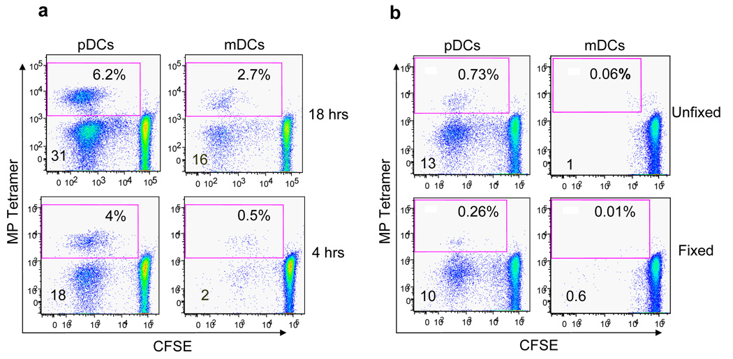
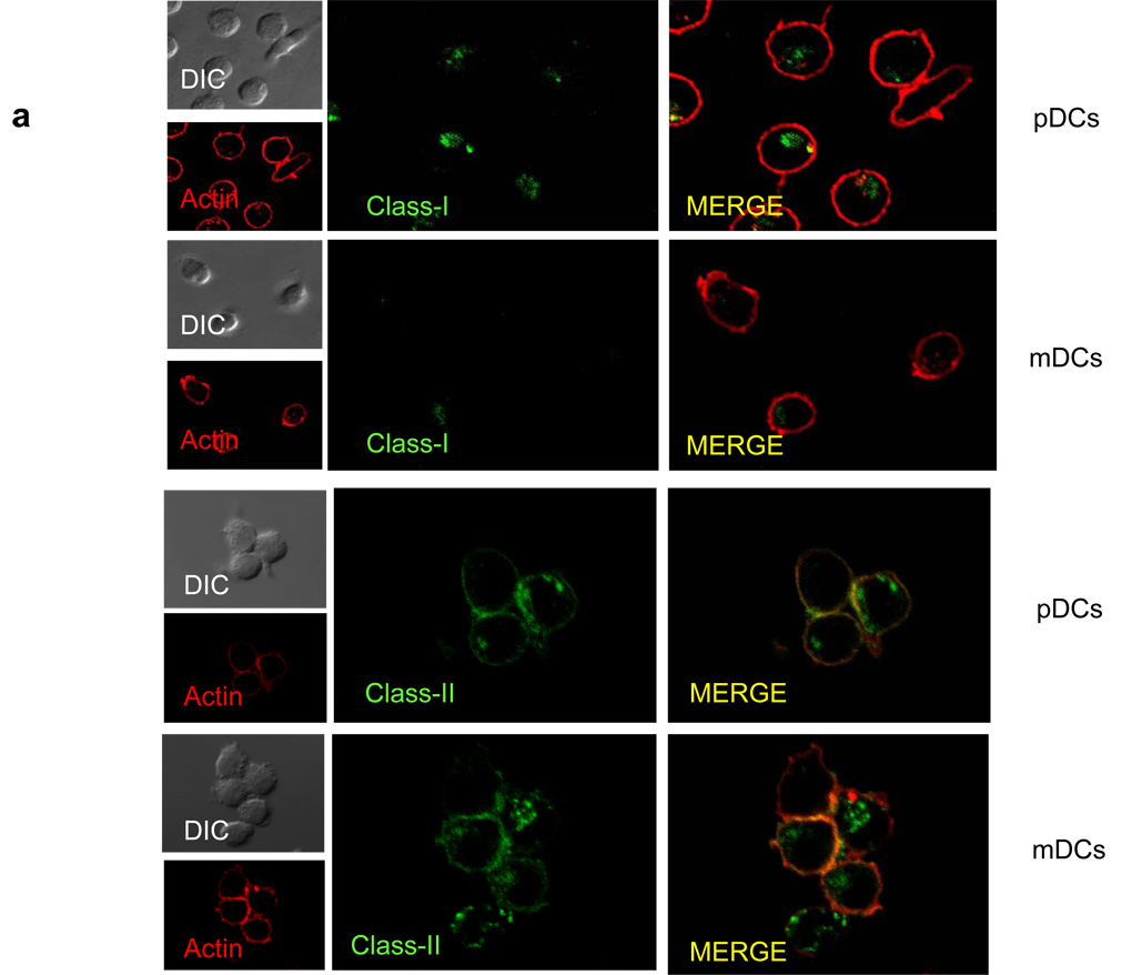
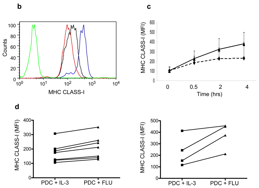
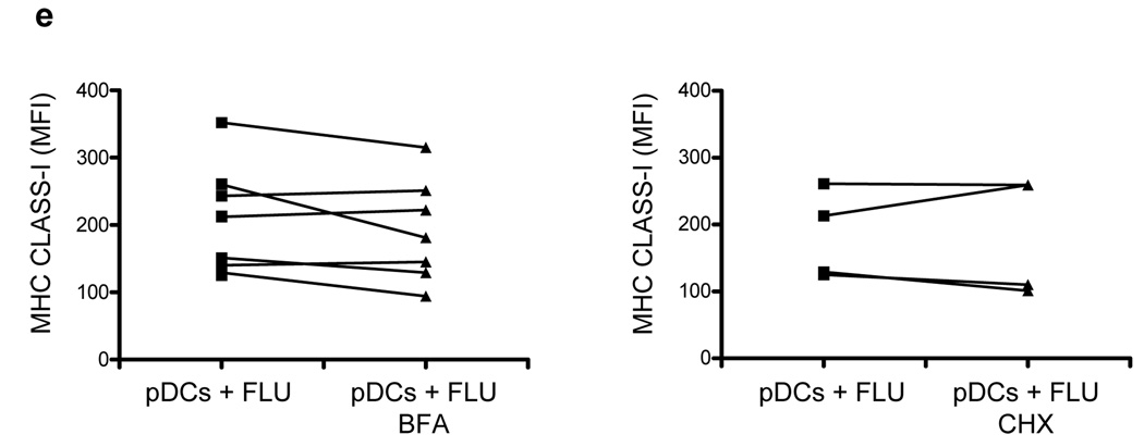
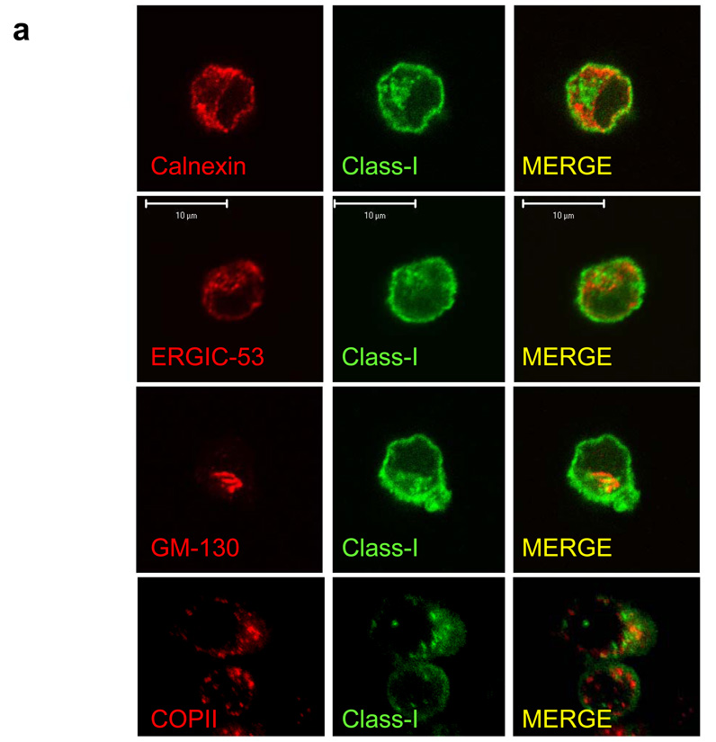
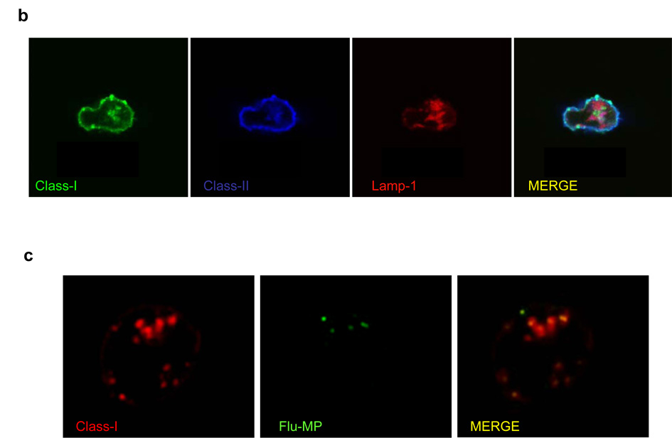
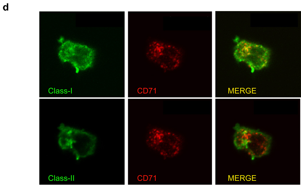
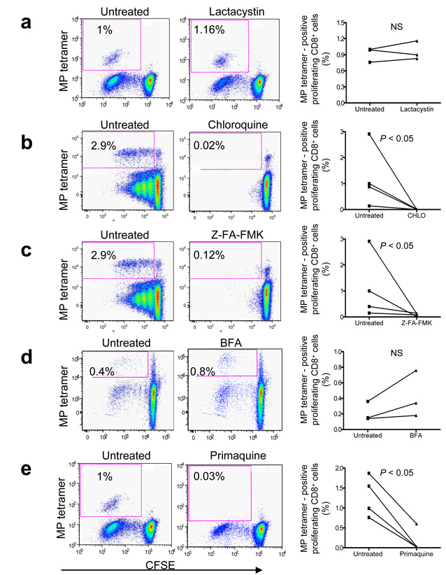
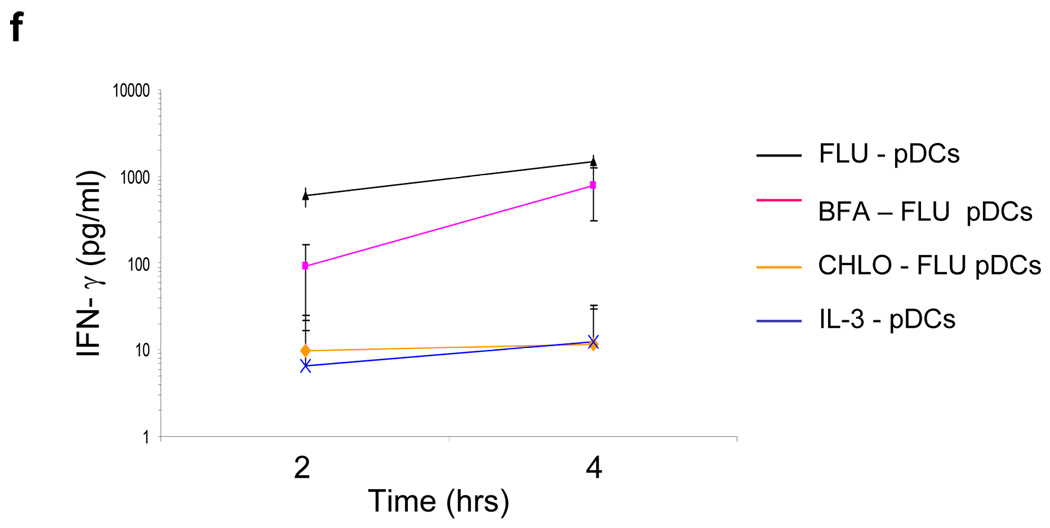
Although plasmacytoid dendritic cells (pDCs) respond to virus replication in a nonspecific way by producing large amounts of type I interferon, a rapid, direct function for pDCs in activating antiviral lymphocytes is less apparent. Here we show that pDCs were able to rapidly initiate antigen-specific antiviral CD8+ T cell responses. After being exposed to virus, pDCs efficiently and rapidly internalized exogenous viral antigens and then presented those antigens on major histocompatibility complex (MHC) class I to CD8+ T cells. Processing of exogenous antigen occurred in endocytic organelles and did not require transit of antigen to the cytosol. Intracellular stores of MHC class I partially localized together with the transferrin receptor and internalized transferrin in endosomes, which suggested that such recycling endosomes are sites for loading peptide onto MHC class I or for peptide transit. Our data demonstrate that pDCs use 'ready-made' stores of MHC class I to rapidly present exogenous antigen to CD8+ T cells.


Figures

Figure 1
Figure 1
Activated pDCs are powerful activators of allogeneic CD8+ T cells.(a) Flow cytometry of CFSE-labeled CD4+ or CD8+ T cells cultured for 6 days with allogenic pDCs and mDCs activated with influenza virus for 18 hrs. Profiles of proliferating CD8+ (upper panels) and CD4+ (bottom panels) T cells are shown. Numbers indicate the percentage of proliferating T cells. (b) Same protocol as in a) in 3 different matched donors. (P = 0.03 n = 4 pDCs vs mDCs/CD8+ T cells) and (P = 0.02, n = 3 pDCs vs mDCs/CD4+ T cells). Data are representative of at least three different matched donors in independent experiments.
Figure 2
Figure 2
pDCs cross-present influenza virus antigen more rapidly and efficiently than mDCs. (a) Flow cytometric profiles of tetramer stained influenza-specific CD8+ T cells activated by pDCs and mDCs exposed to influenza virus for either 18 hrs (upper panels) or 4 hrs (bottom panels). pDCs and mDCs were then cultured for 6 days with autologous CFSE labeled CD8+ T cells. Numbers in the boxes represent percentage of MP tetramer positive proliferating CD8+ T cells. Numbers on left side indicate the percentage of total proliferating CD8+ T cells. Data are representative of three different experiments. (b) Same protocol as in a) except pDCs and mDCs were treated with influenza virus and then fixed prior to 6 day culture with CFSE-labeled CD8+ T cells (bottom panels). Upper panels show the percentage of MP tetramer positive proliferating CD8+ T cells activated by unfixed 6 hr influenza virus treated pDCs and mDCs. Numbers in the boxes indicate percentage of MP-tetramer positive proliferating CD8+ T cells. Numbers on left side indicate the percentage of total proliferating CD8+ T cells. Data are representative of three different matched donors.
Figure 3
Figure 3
MHC class I exists in large perinuclear intracellular pools within the pDCs and rapidly translocates to the cell surface after viral activation of pDCs. (a) Fluorescence microscopy of immature peripheral blood pDCs and mDCs stained with MHC class I (upper two panels) and II (lower two panels) and phalloidin and then analyzed by confocal microscopy. Colocalization of staining was visualized in yellow in merged figures. (b) Flow cytometry of peripheral blood pDCs after influenza virus activation. Sorted pDCs (red line) were incubated in the presence of IL-3 (black line) or influenza virus (blue line) for 4 hrs and stained with anti-MHC class I antibody. Green line shows the isotype control staining. Data are representative of four different donors. (c) Kinetic of MHC class I surface expression on pDCs cultured in the presence of IL-3 (dashed line) and influenza virus (black line). Results are expressed as mean fluorescence intensity (MFI) and represent the mean ±s.d. of four different experiments. (d) Graphs of MHC class I surface expression MFI assessed after either 30 mins (left panel) (n = 7; P = 0.009) or 4 hrs (right panel) (n = 4; P = 0.046) of pDCs culture in the presence of either IL-3 or influenza virus. (e) pDCs were pre-treated with either BFA (right panel, n = 7; P = NS) or CHX (left panel, n = 4; P = NS) and cultured in the presence of influenza virus for additional 30 mins. Data are representative of at least four independent experiments.
Figure 3
Figure 3
MHC class I exists in large perinuclear intracellular pools within the pDCs and rapidly translocates to the cell surface after viral activation of pDCs. (a) Fluorescence microscopy of immature peripheral blood pDCs and mDCs stained with MHC class I (upper two panels) and II (lower two panels) and phalloidin and then analyzed by confocal microscopy. Colocalization of staining was visualized in yellow in merged figures. (b) Flow cytometry of peripheral blood pDCs after influenza virus activation. Sorted pDCs (red line) were incubated in the presence of IL-3 (black line) or influenza virus (blue line) for 4 hrs and stained with anti-MHC class I antibody. Green line shows the isotype control staining. Data are representative of four different donors. (c) Kinetic of MHC class I surface expression on pDCs cultured in the presence of IL-3 (dashed line) and influenza virus (black line). Results are expressed as mean fluorescence intensity (MFI) and represent the mean ±s.d. of four different experiments. (d) Graphs of MHC class I surface expression MFI assessed after either 30 mins (left panel) (n = 7; P = 0.009) or 4 hrs (right panel) (n = 4; P = 0.046) of pDCs culture in the presence of either IL-3 or influenza virus. (e) pDCs were pre-treated with either BFA (right panel, n = 7; P = NS) or CHX (left panel, n = 4; P = NS) and cultured in the presence of influenza virus for additional 30 mins. Data are representative of at least four independent experiments.
Figure 4
Figure 4
MHC class I is not localized in the traditional ER-Golgi transport pathway within pDCs. (a–d) Confocal fluorescence microscopy. (a) Colocalization of calnexin, ERGIC-53, COP-II and GM-130 with MHC class I in immature pDCs. (b) Colocalization of MHC class I with MHC class-II and LAMP-1 in immature pDCs analyzed. (c) Immature peripheral blood pDCs were exposed to influenza virus for 45 min, fixed and stained with antibodies to Flu-MP, and MHC class I. (d) MHC class I colocalizes with CD71. Immature pDCs were fixed, permeabilized and stained for MHC class I, MHC class II and CD71 (upper and bottom panels, respectively). Data are representative of three independent experiments.
Figure 4
Figure 4
MHC class I is not localized in the traditional ER-Golgi transport pathway within pDCs. (a–d) Confocal fluorescence microscopy. (a) Colocalization of calnexin, ERGIC-53, COP-II and GM-130 with MHC class I in immature pDCs. (b) Colocalization of MHC class I with MHC class-II and LAMP-1 in immature pDCs analyzed. (c) Immature peripheral blood pDCs were exposed to influenza virus for 45 min, fixed and stained with antibodies to Flu-MP, and MHC class I. (d) MHC class I colocalizes with CD71. Immature pDCs were fixed, permeabilized and stained for MHC class I, MHC class II and CD71 (upper and bottom panels, respectively). Data are representative of three independent experiments.
Figure 5
Figure 5
Intracellular stores of MHC class I are localized in immature pDCs within the recycling endosomal compartment. (a–e) Dot plots show MP-tetramer CD8 proliferation after FLU-fixed pDC stimulation. pDCs are pre-treated with different inhibitors as shown in the correspondent plots and cultured for 6 hours with influenza virus. pDCs were then fixed and cultured for 6 days with CFSE-labeled CD8+ T cells. Left panels (untreated) show MP-tetramer positive CD8+ T cells stimulated with FLU-treated fixed pDCs in the absence of inhibitors. Right panels show results obtained in different matched donors. (f) IFN-γ secretion by MP specific cells after activation by FLU-treated fixed pDCs. MP specific cell lines are cultured with pDCs exposed to influenza virus in presence or absence of chloroquine and BFA inhibitors followed by fixation. As control, MP specific cell lines are stimulated by IL-3 pDCs. IFN-g is measured by Luminex assay. Results are expressed as the mean±s.d. of three independent experiments.
Figure 5
Figure 5
Intracellular stores of MHC class I are localized in immature pDCs within the recycling endosomal compartment. (a–e) Dot plots show MP-tetramer CD8 proliferation after FLU-fixed pDC stimulation. pDCs are pre-treated with different inhibitors as shown in the correspondent plots and cultured for 6 hours with influenza virus. pDCs were then fixed and cultured for 6 days with CFSE-labeled CD8+ T cells. Left panels (untreated) show MP-tetramer positive CD8+ T cells stimulated with FLU-treated fixed pDCs in the absence of inhibitors. Right panels show results obtained in different matched donors. (f) IFN-γ secretion by MP specific cells after activation by FLU-treated fixed pDCs. MP specific cell lines are cultured with pDCs exposed to influenza virus in presence or absence of chloroquine and BFA inhibitors followed by fixation. As control, MP specific cell lines are stimulated by IL-3 pDCs. IFN-g is measured by Luminex assay. Results are expressed as the mean±s.d. of three independent experiments.

Comment in

Similar articles

Cited by

References

    1. Banchereau J, Steinman RM. Dendritic cells and the control of immunity. Nature. 1998;392:245–252. - PubMed
    1. Shortman K, Liu YJ. Mouse and human dendritic cell subtypes. Nat. Rev. Immunol. 2002;2:151–161. - PubMed
    1. Mellman I, Steinman RM. Dendritic cells: specialized and regulated antigen processing machines. Cell. 2001;106:255–258. - PubMed
    1. Reis ESousa. Dendritic cells in a mature age. Nat. Rev. Immunol. 2006;6:476–483. - PubMed
    1. Watts C. The exogenous pathway for antigen presentation on major histocompatibility complex class II and CD1 molecules. Nat. Immunol. 2004;5:685–692. - PubMed

Publication types

MeSH terms

Substances