Metabolite induction of Caenorhabditis elegans dauer larvae arises via transport in the pharynx
- PMID: 18376812
- PMCID: PMC2692194
- DOI: 10.1021/cb700269e
Metabolite induction of Caenorhabditis elegans dauer larvae arises via transport in the pharynx
Abstract
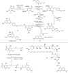
Caenorhabditis elegans sense natural chemicals in their environment and use them as cues to regulate their development. This investigation probes the mechanism of sensory trafficking by evaluating the processing of fluorescent derivatives of natural products in C. elegans. Fluorescent analogs of daumone, an ascaroside, and apigenin were prepared by total synthesis and evaluated for their ability to induce entry into a nonaging dauer state. Fluorescent imaging detailed the uptake and localization of every labeled compound at each stage of the C. elegans life cycle. Comparative analyses against natural products that did not induce dauer indicated that dauer-triggering natural products accumulated in the cuticle of the pharnyx. Subsequent transport of these molecules to amphid neurons signaled entry into the dauer state. These studies provide cogent evidence supporting the roles of the glycosylated fatty acid daumone and related ascarosides and the ubiquitous plant flavone apigenin as chemical cues regulating C. elegans development.
Figures


Similar articles
-
Effects of a Caenorhabditis elegans dauer pheromone ascaroside on physiology and signal transduction pathways.J Chem Ecol. 2009 Feb;35(2):272-9. doi: 10.1007/s10886-009-9599-3. Epub 2009 Feb 4. J Chem Ecol. 2009. PMID: 19190963
-
Chemical structure and biological activity of the Caenorhabditis elegans dauer-inducing pheromone.Nature. 2005 Feb 3;433(7025):541-5. doi: 10.1038/nature03201. Nature. 2005. PMID: 15690045
-
Ascaroside activity in Caenorhabditis elegans is highly dependent on chemical structure.Bioorg Med Chem. 2013 Sep 15;21(18):5754-69. doi: 10.1016/j.bmc.2013.07.018. Epub 2013 Jul 18. Bioorg Med Chem. 2013. PMID: 23920482 Free PMC article.
-
[Genetics and evolution of developmental plasticity in the nematode C. elegans: Environmental induction of the dauer stage].Biol Aujourdhui. 2020;214(1-2):45-53. doi: 10.1051/jbio/2020006. Epub 2020 Aug 10. Biol Aujourdhui. 2020. PMID: 32773029 Review. French.
-
Methods for evaluating the Caenorhabditis elegans dauer state: standard dauer-formation assay using synthetic daumones and proteomic analysis of O-GlcNAc modifications.Methods Cell Biol. 2011;106:445-60. doi: 10.1016/B978-0-12-544172-8.00016-5. Methods Cell Biol. 2011. PMID: 22118287 Review.
Cited by
-
The marginal cells of the Caenorhabditis elegans pharynx scavenge cholesterol and other hydrophobic small molecules.Nat Commun. 2019 Sep 2;10(1):3938. doi: 10.1038/s41467-019-11908-0. Nat Commun. 2019. PMID: 31477732 Free PMC article.
-
Effects of a Caenorhabditis elegans dauer pheromone ascaroside on physiology and signal transduction pathways.J Chem Ecol. 2009 Feb;35(2):272-9. doi: 10.1007/s10886-009-9599-3. Epub 2009 Feb 4. J Chem Ecol. 2009. PMID: 19190963
-
Molecular time-course and the metabolic basis of entry into dauer in Caenorhabditis elegans.PLoS One. 2009;4(1):e4162. doi: 10.1371/journal.pone.0004162. Epub 2009 Jan 8. PLoS One. 2009. PMID: 19129915 Free PMC article.
-
Caenorhabditis elegans pheromones regulate multiple complex behaviors.Curr Opin Neurobiol. 2009 Aug;19(4):378-88. doi: 10.1016/j.conb.2009.07.007. Epub 2009 Aug 7. Curr Opin Neurobiol. 2009. PMID: 19665885 Free PMC article. Review.
-
De novo GTP biosynthesis is critical for virulence of the fungal pathogen Cryptococcus neoformans.PLoS Pathog. 2012;8(10):e1002957. doi: 10.1371/journal.ppat.1002957. Epub 2012 Oct 11. PLoS Pathog. 2012. PMID: 23071437 Free PMC article.
References
-
- Lee DL. In: The Biology of Nematodes. 1st ed. Lee DL, editor. Taylor & Francis; New York: 2002. pp. 61–73.
-
- Moran NA. Symbiosis. Curr. Biol. 2006;16:R866–R871. - PubMed
-
- Golden JW, Riddle DL. A pheromone influences larval development in the nematode Caenorhabditis elegans. Science. 1982;218:578–580. - PubMed
Publication types
MeSH terms
Substances
Grants and funding
LinkOut - more resources
Full Text Sources
Other Literature Sources

