. 2008 May 15;75(10):1946-58.
doi: 10.1016/j.bcp.2008.02.016. Epub 2008 Mar 10.

Quercetin augments TRAIL-induced apoptotic death: involvement of the ERK signal transduction pathway

Affiliations

Quercetin augments TRAIL-induced apoptotic death: involvement of the ERK signal transduction pathway

Young-Ho Kim et al. Biochem Pharmacol. .

Abstract

Combined treatment with quercetin and TRAIL induced cytotoxicity and enhanced annexin V staining and poly (ADP-ribose) polymerase (PARP) cleavage in human prostate cancer cell lines DU-145 and PC-3. These indicators of apoptosis resulted from the activation of caspase-8, -9, and -3. Although the expression levels of FLIPs, cIAP1, cIAP2, and the Bcl-2 family were not changed in quercetin-treated cells, significant downregulation of survivin occurred. Knockdown survivin by siRNA significantly increased TRAIL-induced apoptosis. We hypothesized that quercetin-induced activation of MAPK (ERK, p38, JNK) is responsible for downregulation of survivin gene expression. To test this hypothesis, we selectively inhibited MAPK during treatment with quercetin. Our data demonstrated that inhibitor of ERK (PD98059), but not p38 MAPK (SB203580) or JNK (SP600125), significantly maintained the intracellular level of survivin during treatment with quercetin. Interestingly, PD98059 also prevented quercetin-induced deacetylation of histone H3. Data from survivin promoter activity assay suggest that the Sp1 transcription factor binds to the survivin promoter region and quercetin inhibits its binding activity through deacetylation of histone H3. Quercetin-induced activation of the ERK-MSK1 signal transduction pathway may be responsible for deacetylation of histone H3. Taken together, our findings suggest that quercetin enhances TRAIL induced apoptosis by inhibition of survivin expression, through ERK-MSK1-mediated deacetylation of H3.


Figures

Fig. 1
Fig. 1. Effect of quercetin on TRAIL-induced cytotoxicity in human prostate DU-145 and PC-3 cells
(A) DU-145 cells were treated for 24 h with quercetin (10-200 μM). (B) DU-145 cells were treated for 24 h with various concentrations of TRAIL (10-100 ng/ml) in the presence or absence of 100 μM quercetin. Cell survival was determined by the trypan blue exclusion assay. Error bars represent standard error from the mean (SEM) for three separate experiments. (C) Cells were treated for 4h with TRAIL (50 ng/ml) in the presence or absence of 100 μM quercetin. After treatment, apoptosis was detected by the FACS analysis. Con: control, T50: TRAIL 50ng/ml, Q100: Quercetin 100 μM, T50+Q: TRAIL 50 ng/ml plus Quercetin 100 μM. PC-3 cells were treated with various concentrations of quercetin (10-100 μM) for 24 h in the presence or absence of 50 ng/ml of TRAIL. The morphological features were analyzed with a phase-contrast microscope (200×) (D) and cell survival was determined by the trypan blue exclusion assay (E). Error bars represent standard error from the mean (SEM) for three separate experiments.
Fig. 2
Fig. 2. Effect of quercetin on TRAIL-induced proteolytic cleavage of PARP and activation of caspases in DU-145 cells
Cells were treated for 24 h with various concentrations of quercetin in the presence or absence of 50 ng/ml TRAIL and then harvested. Cell lysates containing equal amounts of protein (20 μg) were separated by SDS-PAGE and immunoblotted with anti-caspase-8, anti-capase-9, anti-caspase-3, or anti-PARP antibody. Actin was used to confirm the amount of proteins loaded in each lane.
Fig. 3
Fig. 3. Intracellular levels of FLIPs, Bcl-2 family, and IAP family proteins during treatment with quercetin in the presence or absence of TRAIL
DU-145 cells were treated for 24 h with various concentrations of quercetin (10-100 μM) in the presence or absence of 50 ng/ml TRAIL. Equal amounts of protein (20 μg) were separated by SDS-PAGE and immunoblotted as described in materials and methods. Protein levels of FLIPs and Bcl-2 family (A) or IAP family (B) were analyzed during treatment with quercetin in the presence or absence of TRAIL. (C) PC-3 cells were treated for 24 h with indicated amount of quercetin (10-100 μM) and survivin level was measured by western blot analysis.
Fig. 4
Fig. 4. Effect of silenced survivin expression on TRAIL-induced apoptosis in DU-145 cells
Cells were transfected with a control vector or silenced survivin oligonucleotide DNA for 24 h. Transfected cells were treated for 4 h with various concentrations of TRAIL (10-200 ng/ml). (A) Cell lysates were subjected to immunoblotting for anti-PARP or anti-survivin. Actin was shown as an internal standard. (B) Cell survival was determined by the trypan blue exclusion assay. Error bars represent standard error from the mean (SEM) for three separate experiments.
Fig. 5
Fig. 5. Effect of quercetin on the mRNA levels, the promoter activity of Survivin and Sp1 binding activity in DU-145 cells
(A) DU-145 cells were treated with the indicated concentration of quercetin. Total RNA was isolated and RT-PCR analysis was performed. A representative study is shown; two additional experiments yielded similar results. (B) Schematic structure of survivin promoter constructs used for testing luciferase activity. (C) Various promoter plasmids were transfected, and treated with quercetin. The cells were lysed and luciferase activity measured.
Fig. 6
Fig. 6. Effect of various MAP kinase or inhibitors on quercetin mediated down-regulation of survivin expression
Quercetin affects the levels of phosphorylated mitogen-activated protein kinase. DU-145 cells were incubated with 100 μM of quercetin for indicated time (A) or with indicated amount of quercetin for 1 h (B) and the levels of phospho-ERK, -JNK, and –p38 were determined by western blotting with phosphor-specific antibodies. DU-145 cells were stimulated with quercetin (100 μM) in the presence or absence of several inhibitors: (C) PD98059 (a specific ERK inhibitor), (D) SB203580 (a specific p38 inhibitor), and (E) SP600125 (a specific JNK inhibitor) as indicated for 24 h and the levels of survivin were determined by western blotting. Protein expression levels of actin in cell lysates were used to confirm the amount of proteins loaded in each lane. (F) DU-145 cells were treated with quercetin (100 μM) and TRAIL (50 ng/ml) in the presence of indicated amount of PD98059 for 24 h. Cell survival was determined by the trypan blue exclusion assay. Error bars represent standard error from the mean (SEM) for three separate experiments.
Fig. 7
Fig. 7. Effect of quercetin on acetylation of histone acetylation and HDAC activity
DU-145 cells were treated with 100 μM (A) or indicated amount (B) of quercetin for 24 h and total level and acetylation of histone H3 and H4 was analyzed. (C) PC-3 cells were treated with indicated amount of quercetin for 24 h and total level and acetylation of histone H3 and H4 was analyzed. (D) DU-145 cells were treated with 100 μM of quercetin for the indicated time and acetylation of histone H3 4 was analyzed. (E) DU-145 cells were treated with quercetin (100 μM) in the presence or absence of several inhibitors: PD98059 (a specific ERK inhibitor), SB203580 (a specific p38 inhibitor), and SP600125 (a specific JNK inhibitor) as indicated for 24 h and the levels of survivin and acetyl H3 were determined by western blotting. (F) HDAC enzyme activity was measured after 24 h exposure to 100 μM of quercetin. Representative results from three independent experiments carried out are shown. NC: negative control, Con: control.
Fig. 8
Fig. 8. Sp1 is associated with quercetin-mediated down-regulation of survivin
(A) DU-145 cells were incubated for the periods indicated in the presence or absence of quercetin. The intracellular levels of Sp1 were determined by western blot analysis. (B) Effect of ectopic expression of Sp1 on survivin expression. DU-145 cells were transfected with various concentrations of Sp1 cDNA plasmid (0.1-1 μg). The expression levels of Sp1 and survivin protein in transfected cells were determined by western blot analysis. (C) Chromatin immunoprecipitation analyses were performed with anti-Sp1 antibody, as described in material and methods. The chromatins were extracted from sonicated cells (Input), protein A without antibody (-), protein A with antibody (anti-Sp1) with/without quercetin treatment, or with pre-immune serum (anti-IgG). Specific promoter regions of the survivin gene were amplified by PCR, separated on 1.2% agarose gels, and stained with ethidium bromide. Also indicated is the relative position of the PCR product generated in ChIP assay.
Fig. 9
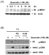
Fig. 9. Effect of quercetin on ERK-MSK1 signal pathway
(A) DU-145 cells were incubated for the periods indicated in the presence or absence of quercetin. Intracellular levels of phospho-MSK1 were determined by western blot analysis. Cells were treated for 1 h (B) and 24 h (C) with 100 μM of quercetin in the absence or presence of the following inhibitors: PD98059 (50 μM), H-89 (20 μM). Immunoblotting was performed using an antibody against phospho-MSK1, phospho-ERK, survivin, or ERK.

Similar articles

Cited by

References

    1. Kelley SK, Ashkenazi A. Targeting death receptors in cancer with Apo2L/TRAIL. Curr Opin Pharmacol. 2004;4:333–9. - PubMed
    1. Thomas LR, Henson A, Reed JC, Salsbury FR, Thorburn A. Direct binding of Fas-associated death domain (FADD) to the tumor necrosis factor-related apoptosis-inducing ligand receptor DR5 is regulated by the death effector domain of FADD. J Biol Chem. 2004 Jul 30;279:32780–5. - PubMed
    1. Rowinsky EK. Targeted induction of apoptosis in cancer management: the emerging role of tumor necrosis factor-related apoptosis-inducing ligand receptor activating agents. J Clin Oncol. 2005;23:9394–407. - PubMed
    1. Block G, Patterson B, Subar A. Fruit, vegetables, and cancer prevention: a review of the epidemiological evidence. Nutr Cancer. 1992;18:1–29. - PubMed
    1. Agullo G, Gamet-Payrastre L, Manenti S, Viala C, Remesy C, Chap H, Payrastre B. Relationship between flavonoid structure and inhibition of phosphatidylinositol 3-kinase: a comparison with tyrosine kinase and protein kinase C inhibition. Biochem Pharmacol. 1997;53:1649–57. - PubMed

Publication types

MeSH terms

Substances