. 2008 Jun;29(17):2565-72.
doi: 10.1016/j.biomaterials.2008.02.023.

A thermoresponsive, microtextured substrate for cell sheet engineering with defined structural organization

Affiliations

A thermoresponsive, microtextured substrate for cell sheet engineering with defined structural organization

Brett C Isenberg et al. Biomaterials. 2008 Jun.

Abstract

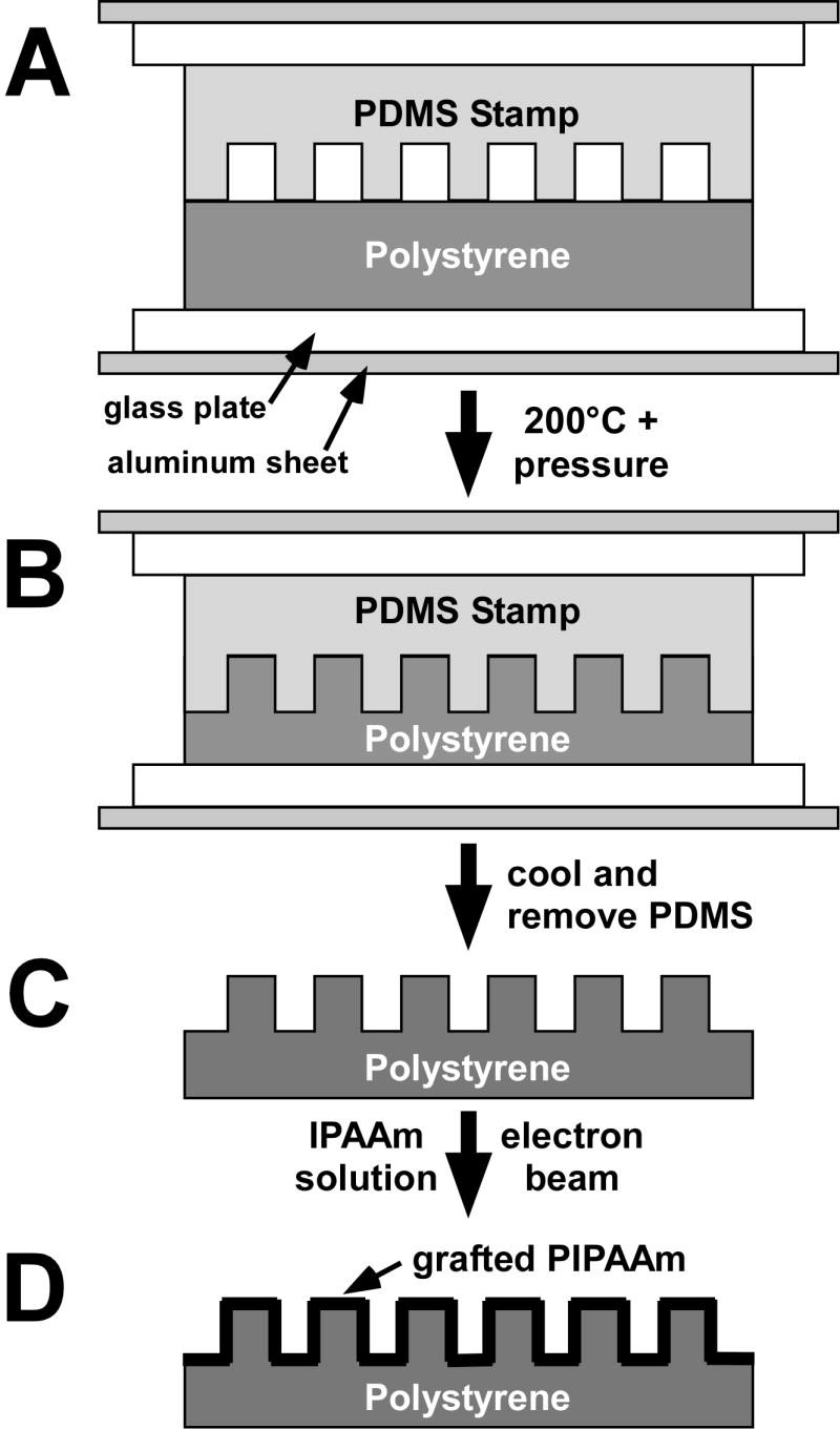
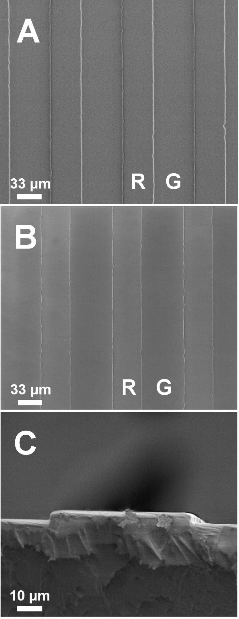
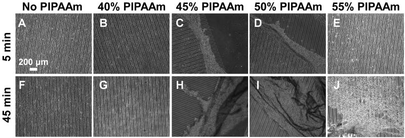
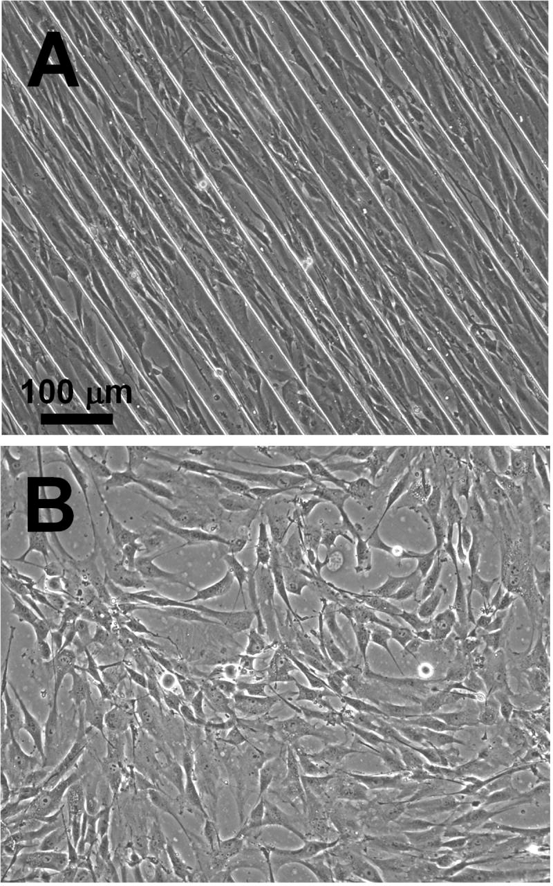
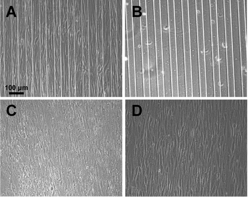
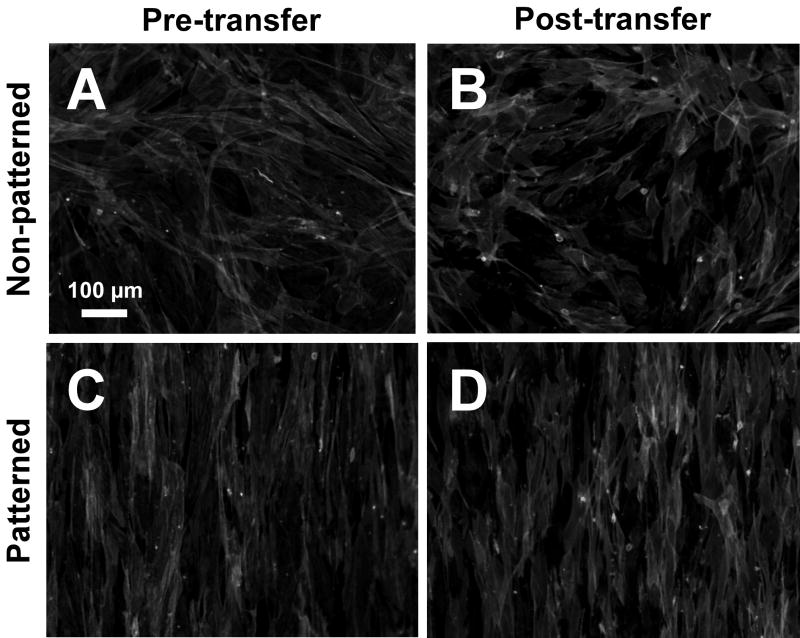
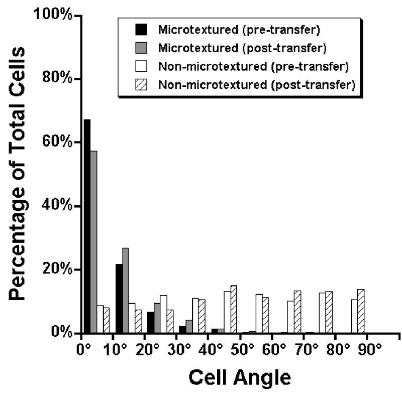
The proper function of many tissues depends critically on the structural organization of the cells and matrix of which they are comprised. Therefore, in order to engineer functional tissue equivalents that closely mimic the unique properties of native tissues it is necessary to develop strategies for reproducing the complex, highly organized structure of these tissues. To this end, we sought to develop a simple method for generating cell sheets that have defined ECM/cell organization using microtextured, thermoresponsive polystyrene substrates to guide cell organization and tissue growth. The patterns consisted of large arrays of alternating grooves and ridges (50 microm wide, 5 microm deep). Vascular smooth muscle cells cultured on these substrates produced intact sheets consisting of cells that exhibited strong alignment in the direction of the micropattern. These sheets could be readily transferred from patterned substrates to non-patterned substrates without the loss of tissue organization. Ultimately, such sheets will be layered to form larger tissues with defined ECM/cell organization that spans multiple length scales.


Figures

Figure 1
Figure 1. Polystyrene substrate microtexturing and PIPAAm grafting
Hot embossing of the PS sheets was accomplished by pressing the microtextured PDMS master molds against a polystyrene sheet between aluminum and glass plates using a custom-built device (A). This device was placed in an oven for 5 minutes at 200°C (B) and subsequently cooled for 5 minutes before removing the PS sheet from the device (C). PS substrates were grafted with PIPAAm by first applying a thin layer of IPAAm dissolved in isopropanol to the surface of the substrate and then exposing them to electron beam irradiation (D). Grafted substrates were washed overnight in deionized water and subsequently dried at 45°C. Pattern dimensions are exaggerated for illustrative purposes.
Figure 2
Figure 2. SEM of microtextured substrates
Scanning electron microscope images of PS substrates show accurate transfer of the microtexture from the PDMS mold (A) to the PS substrate (B). The ridge and groove regions of the substrates are marked with R and G, respectively. Panel C shows a cross-section of a microtextured PS substrate showing the microtexture height is approximately 5 μm.
Figure 3
Figure 3. Thermoresponsive cell sheet detachment is dependent on PIPAAm grafting density
The temperature of the cultures was dropped to 20°C and observed at 5 minute (A-E) and 45 minute (F-J) time points to assess cell detachment. Cell sheets grown on substrates without grafted PIPAAm (A, F) as well as those on 40% PIPAAm substrates (B, G) did not detach from the substrate within 45 minutes. Cell sheets grown on substrates made with 45% and 50% IPAAm began to detach within 5 minutes (C and D, respectively) and could be completely removed by 45 minutes (H and I, respectively). Sheets grown on substrates made with 55% IPAAm began to detach within 45 minutes, but did not detach as intact sheets (E, J).
Figure 4
Figure 4. Cell sheets grown on microtextured, PIPAAm-grafted substrates orient in direction of micropattern
Brightfield images of AoSMCs grown on microtextured PS substrates show that the cells oriented in the direction of the grooves (A), but oriented randomly on non-microtextured substrates (B). The substrates in these images were grafted with 45% PIPAAm.
Figure 5
Figure 5. Transferred cell sheets retain alignment
Cell sheets grown on thermoresponsive, microtextured substrates were transferred to non-textured TCPS culture dishes using a custom-built gelatin-coated manipulator (see text for details) without loss of cell orientation: an organized cell sheet on a microtextured 45% PIPAAm-grafted substrate (A), the microtextured PIPAAm substrate after cell sheet had been removed showing no cells or matrix were left behind after detachment (B), the cell sheet attached to the gelatin-coated manipulator showing that the sheet remains organized following detachment (C), and the cell sheet attached to a non-patterned substrate post-transfer showing retention of cell/matrix organization.
Figure 6
Figure 6. Cell sheet cytoskeletal organization pre- and post-transfer
Cytoskeletal organization of cell sheets was assessed by staining f-actin fibrils with Alexa Fluor 568 Phalloidin (Molecular Probes; 1:200 dilution). (A) Unpatterned, pre-transfer. (B) Unpatterned, post-transfer. (C) Patterned, pre-transfer. (D) Patterned, post-transfer. In panels A and C (pre-transfer), cell sheets were still attached to PIPAAm-grafted PS substrates, while in panels B and D (post-transfer), the cell sheets have been transferred from PIPAAm-grafted substrates to TCPS.
Figure 7
Figure 7. Cell orientation distribution on microtextured and non-microtextured substrates, pre- and post-transfer
Cell orientation was quantified staining the cell nuclei of cells grown on substrates grafted with 45% PIPAAm with Hoechst 33342 for 30 minutes. For each individual substrate, five images were taken at random locations under epifluorescence and the cell orientation was measured by finding the angle between the long axis of the nuclei and the direction of the grooves. An angle of 0° indicated parallel orientation to the grooves, while an angle of 90° indicates a perpendicular orientation to the grooves. For non-microtextured substrates, the angle was arbitrarily chosen for each image. Cells grown on non-microtextured substrates (white bars) showed random distribution of cell orientation angles (no preferential orientation), while cells grown on microtextured substrates (black bars) showed a strong orientation response in the direction of the grooves. For both microtextured and non-microtextured substrates, the distribution of cell orientation angles did not change appreciably following transfer via the gelatin manipulator method (gray bars and hatched bars, respectively).

Similar articles

Cited by

References

    1. Fung YC. Biomechanics: Mechanical Properties of Living Tissues. New York: Spinger-Verlag; 1993.
    1. Isenberg BC, Williams C, Tranquillo RT. Small-diameter artificial arteries engineered in vitro. Circ Res. 2006;98(1):25–35. - PubMed
    1. Teebken OE, Haverich A. Tissue engineering of small diameter vascular grafts. Eur J of Vasc Endovasc Surg. 2002;23(6):475–485. - PubMed
    1. Schmedlen RH, Elbjeirami WM, Gobin AS, West JL. Tissue engineered small-diameter vascular grafts. Clin Plast Surg. 2003;30(4):507–517. - PubMed
    1. Canham PB, Talman EA, Finlay HM, Dixon JG. Medial collagen organization in human arteries of the heart and brain by polarized light microscopy. Connect Tissue Res. 1991;26(12):121–134. - PubMed

Publication types