. 2008 Jun 15;171(1):30-8.
doi: 10.1016/j.jneumeth.2008.01.033. Epub 2008 Mar 10.

Imaging the spread of reversible brain inactivations using fluorescent muscimol

Affiliations

Imaging the spread of reversible brain inactivations using fluorescent muscimol

Timothy A Allen et al. J Neurosci Methods. .

Abstract

Muscimol is a GABA A-agonist that causes rapid and reversible suppression of neurophysiological activity. Interpretations of the effects of muscimol infusions into the brain have been limited because of uncertainty about spread of the drug around the injection site. To solve this problem, the present study explored the use of a fluorophore-conjugated muscimol molecule (FCM). Whole-cell recordings from horizontal brain slices demonstrated that bath-applied FCM acts like muscimol in reversibly suppressing excitatory synaptic transmission. Two types of in vivo experiments demonstrated that the behavioral effects of FCM infusion are similar to the behavioral effects of muscimol infusion. FCM infusion into the rat amygdala before fear conditioning impaired both cued and contextual freezing, which were tested 24 or 48 h later. Normal fear conditioning occurred when these same rats were subsequently given phosphate-buffered saline infusions. FCM infusion into the dorsomedial prefrontal cortex impaired accuracy during a delayed-response task. Histological analysis showed that the region of fluorescence was restricted to 0.5-1mm from the injection site. Myelinated fiber tracts acted as diffusional barriers, thereby shaping the overall spread of fluorescence. The results suggest that FCM is indeed useful for exploring the function of small brain regions.


Figures

Figure 1
Figure 1
Molecular structure of muscimol compared to fluorescent muscimol. A, Structure of muscimol. The molecular formula is C4H6N2O2. The molecular weight is 114.10. B, Structure of Bodipy-TMR-X®-conjugated muscimol (FCM). The molecular formula is C31H36BF2N5O5. The molecular weight is 607.46. The structure of FCM was reproduced with permission from Molecular Probes (Molecular Probes, Carlsbad, CA).
Figure 2
Figure 2
Parallel changes in the excitatory postsynaptic potential (EPSP) amplitude and the input resistance (RN) in a layer V perirhinal neuron. The left ordinate shows EPSP amplitudes, which are plotted as a function of time (open circles connected by a dashed line). The right ordinate shows concurrent input resistance (filled squares connected by a solid line). Each point is the average of ten measurements. Error bars are ± 1 SEM. FCM was bath applied at 20 min (downward arrow). Washout started at 35 min (upward arrow). Infusion with FCM caused a rapid and large reduction in the EPSP amplitudes and a parallel decline in the input resistance. Following washout (upward arrow), the input resistance and EPSP amplitude recovered over the time course of hours.
Figure 3
Figure 3
Infusions of FCM can result in asymmetrical diffusion gradients around the injection site. The example shown here is a 1 μL infusion of FCM into area 36 of perirhinal cortex. A, A coronal slice of the right hemisphere at approximately −4.7 A/P, −6.8 M/L −6.7 D/V of the Paxinos and Watson atlas (1998). The image is an overlay of a dark field image of PBS-moist tissue and the FCM. B, A magnified image of the FCM spread. There is a hint that the spread of fluorescence was impeded by white matter. The checkered box indicates the area analyzed for luminosity. C, Quantification of the luminosity of the FCM. The asterisk to the right of the peak fluorescence indicates the location of damage from the infusion needle. The highest intensity of fluorescence is represented by the white peak at 0.5 mm (x-axis) by 0.0 − 0.2 (y-axis). The dashed line represents the approximate location of deep cerebral white matter medial to the infusion site. The white matter acted as a diffusion barrier, causing a rapid drop in luminosity. Abbreviations: dcw, deep cerebral white matter; MGM, medial geniculate nucleus of the thalamus; HP, hippocampus; TE, temporal cortex; PR, perirhinal cortex; and EC, entorhinal cortex.
Figure 4
Figure 4
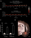
Effect of pre-training infusions of fluorescent muscimol (FCM) into the basolateral nucleus of the amygdala (BLA) on cued and contextual fear conditioning (n = 3). A, Spectrogram of the cue, a 22 kHz ultrasonic vocalization (USV) that was prerecorded from a conspecific. The arrow indicates the time of a 0.4 s, 0.8 mA foot shock that coterminated with the cue. B, During testing, the USV was presented repeatedly for 360 s (60 s is shown). C, Conditioning in rats infused with FCM or vehicle (PBS). Cue conditioning was tested in a shifted context. FCM severely impaired the acquisition of cued and contextual fear conditioning, similar to what has been previously reported using muscimol. Normal fear conditioning occurred after PBS infusions. D, Image of FCM infusion into BLA. The red fluorescence is true color, overlaid with PBS-moist tissue for gross histological localization. The fluorescence is well-restricted to BLA. Calibration bar is 1 mm. Abbreviation: rh, rhinal sulcus.
Figure 5
Figure 5
Performance effects of FCM infusions into the dorsomedial prefrontal cortex (dmPFC) on a delayed-response task (n = 4). The coordinates used for the dmPFC are as follows: AP: +3.2, DV −3.7, ML ± 1.4. Infusions of both muscimol and FCM into dmPFC significantly decreased the number of correct trials. The majority of the errors were premature responses (Naranayan et al., 2006; Naranayan and Laubach, 2006). A, A schematic representation of the trials in the delayed-response task. B, The percentage of correct responses after infusion of saline, muscimol and FCM. C, A sample FCM infusion into the dmPFC. The slice is counterstained with a green fluorescent Nissl (Molecular Probes). D, A false-color plot of the spread of FCM averaged over 8 slices from 4 rats. The images are aligned to the highest-intensity fluorescence in each slice. Fluorescence intensity is normalized within each slice. The spread of fluorescence was 0.5 – 1.0 mm around the injection site.

References

    1. Allen TA, Furtak SC, Brown TH. Single-unit responses to 22 kHz ultrasonic vocalizations in rat perirhinal cortex. Behav Brain Res. 2007;182(2):327–336. - PMC - PubMed
    1. Arikan R, Blake NMJ, Erinjeri JP, Woolsey TA, Giraud L, Highstein SM. A method to measure the effective spread of focally injected muscimol into the central nervous system with electrophysiology and light microscopy J. Neurosci Methods. 2002;118:51–57. - PubMed
    1. Anderson JW. The production of ultrasonic sounds by laboratory rats and other mammals. Science. 1954;19:808–809. - PubMed
    1. Beaumont K, Chilton WS, Yamamura HI, Enna SJ. Muscimol binding in rat brain: Association with synaptic GABA receptors. Brain Res. 1978;148:153–162. - PubMed
    1. Blanchard RJ, Blanchard DC. Crouching as an index of fear. J Comp Physiol Psychol. 1969;67:370–375. - PubMed

Publication types

MeSH terms