. 2008 Sep;34(9):1421-33.
doi: 10.1016/j.ultrasmedbio.2008.01.016. Epub 2008 Apr 18.

Ultrasound-enhanced thrombolysis using Definity as a cavitation nucleation agent

Affiliations

Ultrasound-enhanced thrombolysis using Definity as a cavitation nucleation agent

Saurabh Datta et al. Ultrasound Med Biol. 2008 Sep.

Abstract

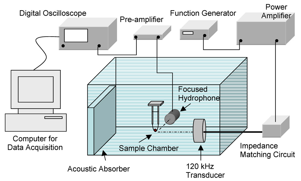
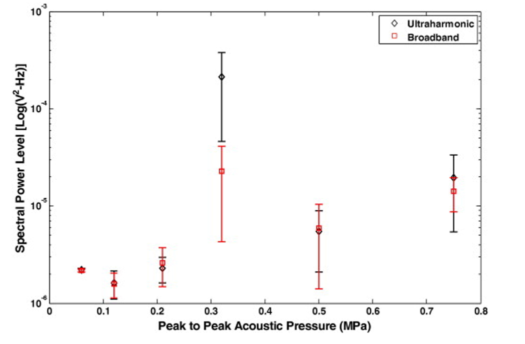
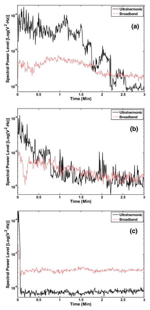
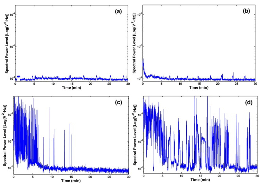
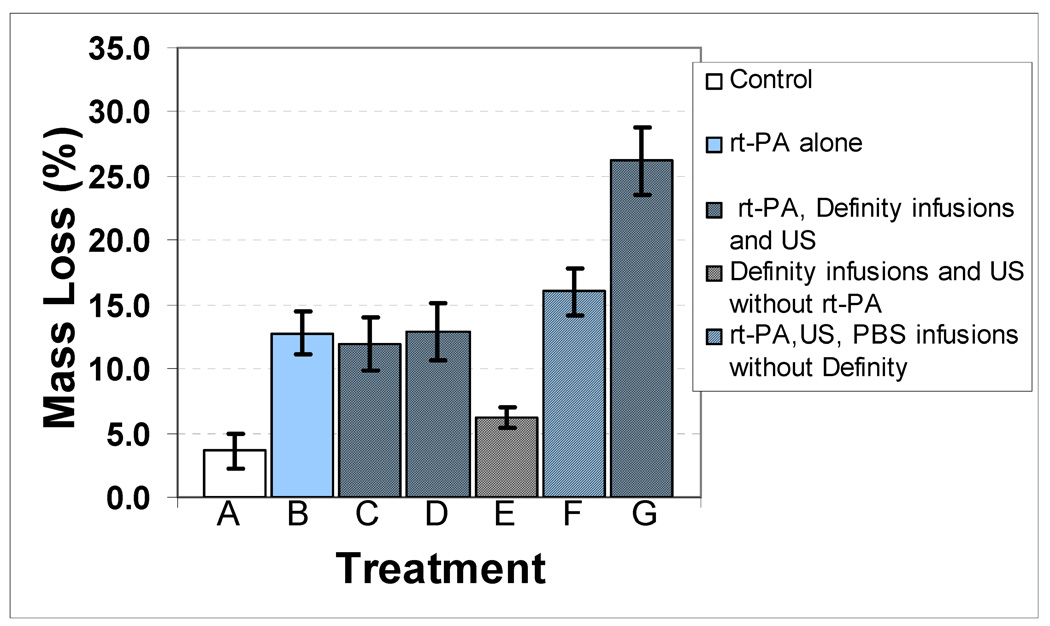
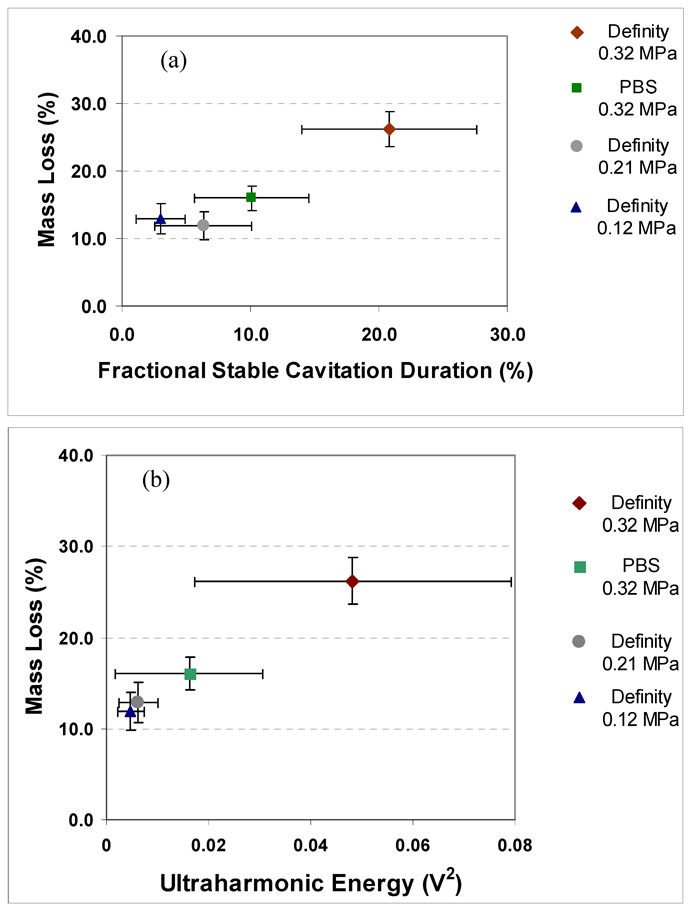
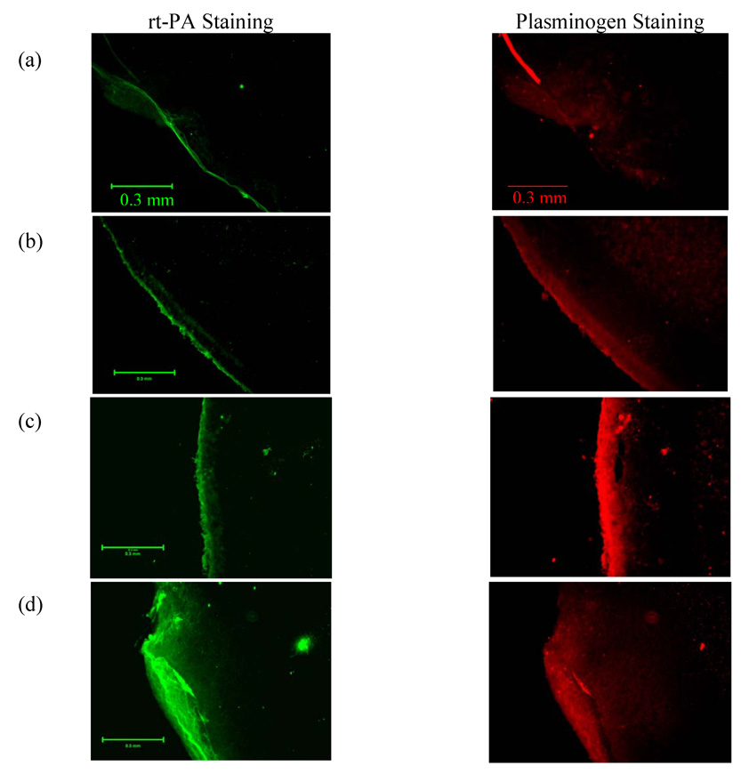
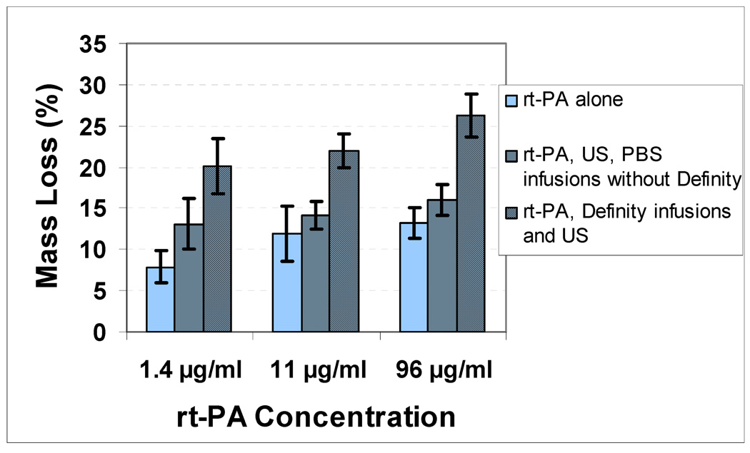
Ultrasound has been shown previously to act synergistically with a thrombolytic agent, such as recombinant tissue plasminogen activator (rt-PA) to accelerate thrombolysis. In this in vitro study, a commercial contrast agent, Definity, was used to promote and sustain the nucleation of cavitation during pulsed ultrasound exposure at 120 kHz. Ultraharmonic signals, broadband emissions and harmonics of the fundamental were measured acoustically by using a focused hydrophone as a passive cavitation detector and used to quantify the level of cavitation activity. Human whole blood clots suspended in human plasma were exposed to a combination of rt-PA, Definity and ultrasound at a range of ultrasound peak-to-peak pressure amplitudes, which were selected to expose clots to various degrees of cavitation activity. Thrombolytic efficacy was determined by measuring clot mass loss before and after the treatment and correlated with the degree of cavitation activity. The penetration depth of rt-PA and plasminogen was also evaluated in the presence of cavitating microbubbles using a dual-antibody fluorescence imaging technique. The largest mass loss (26.2%) was observed for clots treated with 120-kHz ultrasound (0.32-MPa peak-to-peak pressure amplitude), rt-PA and stable cavitation nucleated by Definity. A significant correlation was observed between mass loss and ultraharmonic signals (r = 0.85, p < 0.0001, n = 24). The largest mean penetration depth of rt-PA (222 microm) and plasminogen (241 microm) was observed in the presence of stable cavitation activity. Stable cavitation activity plays an important role in enhancement of thrombolysis and can be monitored to evaluate the efficacy of thrombolytic treatment.


Figures

Figure 1
Figure 1
Experimental setup for ultrasound exposure of human blood clots placed in a sample holder containing plasma and Definity®. A focused hydrophone placed 90 degrees to the acoustic axis of the 120-kHz transducer and with its focus coincident with the blood clot was used as a passive cavitation detector.
Figure 2
Figure 2
Schematic of the signal processing algorithm to determine cavitation dose from scattered acoustic signals received by the broadband focused hydrophone.
Figure 3
Figure 3
Spectral power levels in ultraharmonic and broadband bands as a function of increasing peak-to-peak acoustic pressure amplitude. The scattered signal is received by a hydrophone placed 90 degrees to the source transducer axis with its focus coincident with the region of the sample holder containing a human clot in plasma and Definity®. Three different samples were exposed at each pressure and error bars represent the standard deviation of 60 measurements.
Figure 4
Figure 4
Representative ultraharmonic and broadband signal levels as function of time following a single infusion of Definity® with peak-to-peak US exposure of a) 0.32 MPa, b) 0.50 MPa and c) 0.75 MPa.
Figure 5
Figure 5
A representative example of the spectral power in the ultraharmonic band over the treatment duration; a) with Definity® infusion but no cavitation (0.12 MPa peak-to-peak), b) with Definity® infusion but no cavitation (0.21 MPa peak-to-peak), c) with infusion of PBS alone at a peak-to-peak pressure of 0.32 MPa (note only initial cavitation activity is observed) and d) with infusion of Definity® resulting in relatively sustained cavitation at the same peak-to-peak pressure (0.32 MPa).
Figure 6
Figure 6
Mass loss (%) as a function of peak-to-peak acoustic pressure for different treatments: A) No rt-PA, Definity® or US, which is the control treatment, B) rt-PA alone, C) rt-PA, Definity® infusions and US (0.12 MPa peak-to-peak pressure amplitude), D) rt-PA, Definity® infusions and US (0.21 MPa peak-to-peak pressure amplitude), E) Definity® infusions and US (0.32 MPa peak-to-peak pressure amplitude) without rt-PA treatment, F) rt-PA, PBS infusions without Definity® and US (0.32 MPa peak-to-peak pressure amplitude) and G) rt-PA, Definity® infusions, and US (0.32 MPa peak-to-peak pressure amplitude). A sample size of six was used for each treatment.
Figure 7
Figure 7
Mass loss (%) and cavitation activity relationship as a function of (a) fractional stable cavitation duration which is a fraction of the total time for which the ultraharmonic energy level was above the noise floor and (b) ultraharmonic energy integrated over a 30 min exposure. A sample size of six was used for each treatment and error bars represent standard deviation of the measurements.
Figure 8
Figure 8
Representative examples of fluorescent dual antibody stained images of clot sections. The left column images are stained green for rt-PA by fluorescien isothiocynate (FITC) conjugated secondary antibody. For the same section the right column images show accumulation of plasminogen stained red by Texas red (TRITC) conjugated secondary antibody. Representative clot section images are shown for a) no rt-PA, Definity® or US (the control treatment), b) rt-PA alone treatment, c) rt-PA, PBS infusions and US (0.32 MPa peak-to-peak amplitude) treatment and d) rt-PA, Definity® infusions and US (0.32 MPa peak-to-peak amplitude) treatment.
Figure 9
Figure 9
Effect of Definity® on clot mass loss as a function of rt-PA concentration. The peak-to-peak pressure amplitude used in all US treatments was 0.32 MPa. A significant enhancement over rt-PA alone treatment can be observed at lowest concentration.

Similar articles

Cited by

References

    1. Akiyama M, Ishibashi T, Yamada T, Furuhata H. Low –frequency ultrasound penetrates the cranium and enhances thrombolysis in vitro. Neurosurgery. 1998;43:828–833. - PubMed
    1. Alexandrov AV, Molina CA, Grotta JC, Garmi Z, Ford SR, Alvarez-Sabin J, Montaner J, Saqqur M, Demchuk AM, Moye LA, Hill MD, Wojner AW. Ultrasound-Enhanced Systemic Thrombolysis for Acute Ischemic Stroke. N. Engl. J. of Med. 2004;351(21):2170–2178. - PubMed
    1. Alexandrov AV, Demchuk AM, Burgin WS, Robinson DJ, Grotta JC. Ultrasound-enhanced thrombolysis for acute ischemic stroke: Phase I. Findings of the CLOTBUST trial. J. of Neuroimaging. 2004;14(2):113–117. - PubMed
    1. Behrens S, Daffertshofer M, Spiegel D, Hennerici M. Low-frequency, low-intensity ultrasound accelerates thrombolisis through the skull. Ultrasound Med Biol. 1999;25:269–273. - PubMed
    1. Birnbaum Y, Luo H, Nagai T, Fishbein MC, Peterson TM, Li S, Kricsfeld D, Porter TR, Siegel RJ. Noninvasive in vivo clot dissolution without a thrombolytic drug: Recanalization of thrombosed iliofemoral arteries by transcutaneous ultrasound combined with intravenous infusion of microbubbles. Circulation. 1998;97:130–134. - PubMed

Publication types

MeSH terms