. 2008 Jul;49(7):3231-8.
doi: 10.1167/iovs.08-1754. Epub 2008 Mar 31.

Role of NADPH oxidase and Stat3 in statin-mediated protection against diabetic retinopathy

Affiliations

Role of NADPH oxidase and Stat3 in statin-mediated protection against diabetic retinopathy

Mohamed Al-Shabrawey et al. Invest Ophthalmol Vis Sci. 2008 Jul.

Abstract

Purpose: Inhibitors of 3-hydroxy-3-methylglutaryl CoA reductase (statins) reduce signs of diabetic retinopathy in diabetic patients and animals. Indirect clinical evidence supports the actions of statins in improving cardiovascular function, but the mechanisms of their protective actions in the retina are not understood. Prior studies have implicated oxidative stress and NADPH oxidase-mediated activation of signal transducer and activator of transcription 3 (STAT3) in diabetes-induced increases in expression of vascular endothelial growth factor (VEGF) and intercellular adhesion molecule (ICAM)-1 and breakdown of the blood-retinal barrier (BRB). Because statins are known to be potent antioxidants, the hypothesis for the current study was that the protective effects of statins in preventing diabetic retinopathy involve blockade of diabetes-induced activation of NADPH oxidase and STAT3.

Methods: The hypothesis was tested by experiments in which rats with streptozotocin (STZ)-induced diabetes and retinal endothelial cells maintained in high-glucose medium were treated with simvastatin. Blood-retinal barrier (BRB) function was assayed by determining extravasation of albumin. Oxidative stress was assayed by measuring lipid peroxidation, protein nitration of tyrosine, dihydroethidine oxidation, and chemiluminescence. Immunoprobe techniques were used to determine the levels of NADPH oxidase subunit expression and STAT3 activation.

Results: These studies showed that simvastatin blocks diabetes or high-glucose-induced increases in VEGF and ICAM-1 and preserves the BRB by a process involving blockade of diabetes/high-glucose-induced activation of STAT3 and NADPH oxidase. Statin treatment also prevents diabetes-induced increases in expression of the NADPH oxidase catalytic and subunit NOX2.

Conclusions: These results suggest that simvastatin protects against the early signs of diabetic retinopathy by preventing NADPH oxidase-mediated activation of STAT3.


Figures
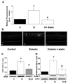
FIGURE 1
FIGURE 1
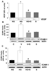
Analysis of the effect of simvastatin (5 mg/kg/d, SC) on diabetes- induced increases in retinal vascular permeability and upregulation of VEGF and ICAM-1 protein in the STZ-induced diabetic rat retina. (A) Rats were injected intravenously with Alexa-Fluor 488-BSA (100 mg/kg), and permeability was determined by morphometric analysis of fluorescence intensity in serial sections. Diabetic retinas had a threefold increase in fluorescence compared with the control (C). Treatment of diabetic rats with simvastatin blocked the permeability increase. (B) Representative images showing the distribution of VEGF immunostaining. VEGF immunoreactivity is concentrated in glial cell processes within the inner retina (arrows). (C) Western blot analysis of VEGF protein showed a significant increase in VEGF expression in the diabetic retina that was blocked by simvastatin. (D) Western blot analysis of ICAM-1 protein showed a five- to sixfold increase in ICAM-1 expression in the diabetic retina that was blocked by simvastatin. Data represent the mean ± SEM of six in each group. *P < 0.01 vs. control; #P < 0.01 vs. diabetic. C, control; D, diabetic; S, simvastatin.
FIGURE 2
FIGURE 2
Measurement of VEGF and ICAM-1 protein levels in BRECs treated with high glucose (25 mM d-glucose, 3 days) or VEGF (20 ng/mL, 24 hours) with or without simvastatin (10 µM) versus control medium (5.5 mM d-glucose). Western blot analyses showed that the high glucose-induced increase in VEGF (A) and ICAM-1 (B) expression was blocked by simvastatin treatment. (C) Western blot analysis shows that the VEGF-induced increase in ICAM-1 expression was blocked by simvastatin treatment. Data represent the mean ± SEM of three to four experiments. *P < 0.01 vs. control, #P < 0.01 vs. high glucose. C, control; HG, high glucose; S, simvastatin; V, VEGF.
FIGURE 3
FIGURE 3
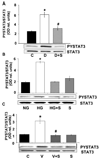
Analysis of STAT3 activation in the diabetic retina and high glucose-treated BRECs. (A) Western blot analysis of STAT3 activation as shown by phosphorylation of tyrosine 705 demonstrates that diabetes caused significant activation of STAT3 compared with the control. This effect was blocked by simvastatin (5 mg/kg/d, SC). Data represent the mean ± SEM for six animals. (B) Western blot analysis showing that high glucose treatment of BRECs (25 mM d-glucose, 72 hours) increased STAT3 activation above the control levels (5.5 mM d-glucose) and that simvastatin (10 µM) blocked the effect. (C) Western blot analysis showing that VEGF treatment of BRECs (20 ng/mL, 24 hours) increased STAT3 activation above the control levels and that simvastatin (10 µM) blocked the effect. Data represent the mean ± SEM of three experiments. *P < 0.01 vs. control, #P < 0.01 vs. high glucose. C, control; HG, high glucose, V, VEGF; S, simvastatin.
FIGURE 4
FIGURE 4
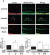
Analysis of NOX2 (gp91phox) and p47phox in control, diabetic, and simvastatin-treated diabetic (5 mg/kg/d, SC) retinas. (A) Retinal sections from control, diabetic, and statin-treated diabetic retinas were reacted with GSI lectin to label the vessels (red) and gp91phox antibody to label NOX2 (green). Merged images show low levels of NOX2 in the normal retina and marked increases in the diabetic retina, especially within the retinal vessels (yellow). (B) Western blot analysis showed a significant increase in NOX2 protein expression in the diabetic retinas compared with the control, which was blocked by statin treatment. (C) Western blot analysis shows increased levels of p47phox protein in the diabetic retina which was inhibited by statin treatment. Data represent the mean ± SEM of six animals/group. *P < 0.01 vs. control, #P < 0.01 vs. diabetic. C, control; D, diabetic; S, simvastatin.
FIGURE 5
FIGURE 5
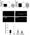
Analysis of ROS formation in the diabetic retina and high glucose-treated BRECs. (A) Levels of lipid peroxides in control, diabetic, and simvastatin-treated (5 mg/kg/d, SC) diabetic retinas as determined by the amount of thiobarbituric acid reactivity with malondialdehyde shows a significant increase in formation of lipid peroxides in the diabetic retina that was blocked by the statin treatment. Data represent the mean ± SEM of six animals/group. *P < 0.01 vs. untreated control, #P < 0.01 vs. diabetic. (B) Analysis of tyrosine nitration in control, diabetic, and simvastatin-treated (5 mg/kg/d) diabetic retinas shows a significant increase in peroxynitrite formation in the diabetic retina that was blocked by the statin treatment. Data represent the mean ± SEM of six animals/group. *P < 0.01 vs. untreated control, #P < 0.01 vs. diabetic. (C) Superoxide generation in the diabetic retina as determined by imaging of the oxidative fluorescent dye DHE in retinal sections in vitro shows a marked increase in the diabetic retinas that was blocked by the statin treatment. The DHE reaction was reduced in retinal sections treated with PEG-SOD, demonstrating specificity of the reaction for superoxide. (D) Analysis of superoxide generation in BRECs treated with high glucose (25 mM d-glucose, 5 days) using chemiluminescence showed a significant increase compared with control cultures treated with normal glucose media (5.5 mM d-glucose). The high-glucose effect was blocked by simvastatin (10 µM) or apocynin (10 mM). Blockade of the chemiluminescence reaction with apocynin demonstrated specificity of the reaction for NADPH oxidase. Data represent the mean ± SEM of four samples/group. *P < 0.01 vs. control, #P < 0.01 vs. high glucose. C, control; D, diabetic; S, simvastatin; NG, normal glucose; HG, high glucose; APO, apocynin.

Similar articles

Cited by

References

    1. Antonetti DA, Lieth E, Barber AJ, Gardner TW. Molecular mechanisms of vascular permeability in diabetic retinopathy. Semin Ophthalmol. 1999;14:240–248. - PubMed
    1. Caldwell RB, Bartoli M, Behzadian MA, et al. Vascular endothelial growth factor and diabetic retinopathy: role of oxidative stress. Curr Drug Targets. 2005;6:511–524. - PubMed
    1. Obrosova IG, Minchenko AG, Marinescu V, et al. Antioxidants attenuate early up regulation of retinal vascular endothelial growth factor in streptozotocin-diabetic rats. Diabetologia. 2001;44:1102–1110. - PubMed
    1. El-Remessy AB, Behzadian MA, Abou-Mohamed G, Franklin T, Cald-well RW, Caldwell RB. Experimental diabetes causes breakdown of the blood-retina barrier by a mechanism involving tyrosine nitration and increases in expression of vascular endothelial growth factor and urokinase plasminogen activator receptor. Am J Pathol. 2003;162:1995–2004. - PMC - PubMed
    1. El-Remessy AB, Al-Shabrawey M, Khalifa Y, Tsai NT, Caldwell RB, Liou GI. Neuroprotective and blood-retinal barrier-preserving effects of cannabidiol in experimental diabetes. Am J Pathol. 2006;168:235–244. - PMC - PubMed

Publication types

MeSH terms