. 2008 May 23;283(21):14717-27.
doi: 10.1074/jbc.M801070200. Epub 2008 Mar 31.

Major histocompatibility complex class II-peptide complexes internalize using a clathrin- and dynamin-independent endocytosis pathway

Affiliations

Major histocompatibility complex class II-peptide complexes internalize using a clathrin- and dynamin-independent endocytosis pathway

Even Walseng et al. J Biol Chem. .

Abstract

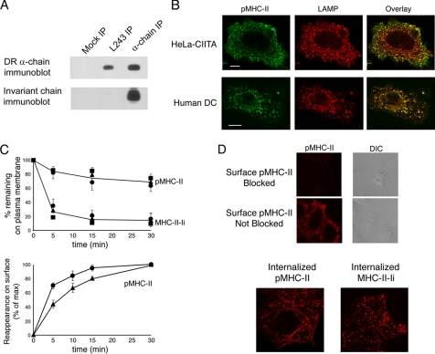
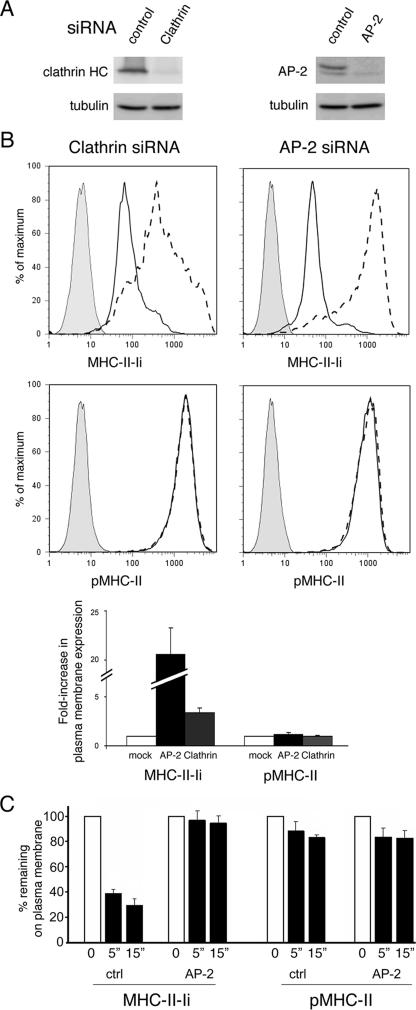
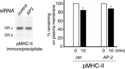
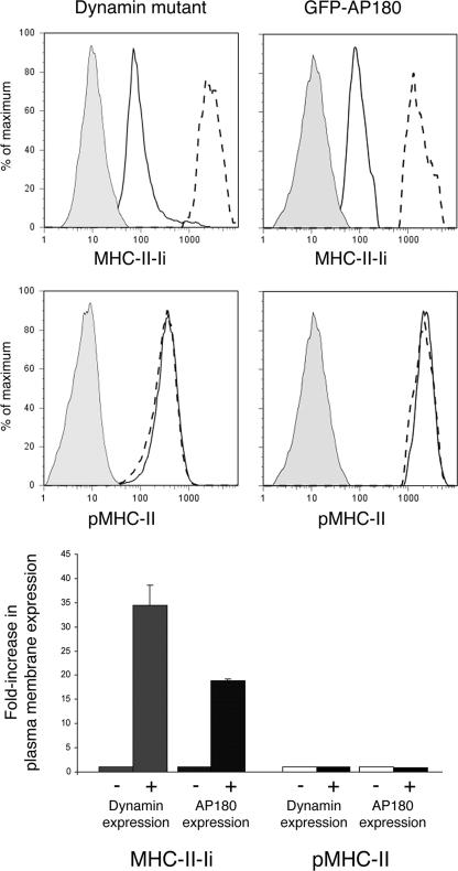
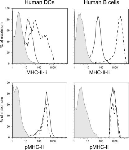
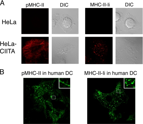
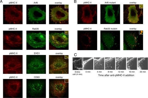
Major histocompatibility complex (MHC) class II molecules (MHC-II) function by binding antigenic peptides and displaying these peptides on the surface of antigen presenting cells (APCs) for recognition by peptide-MHC-II (pMHC-II)-specific CD4 T cells. It is known that cell surface MHC-II can internalize, exchange antigenic peptides in endosomes, and rapidly recycle back to the plasma membrane; however, the molecular machinery and trafficking pathways utilized by internalizing/recycling MHC-II have not been identified. We now demonstrate that unlike newly synthesized invariant chain-associated MHC-II, mature cell surface pMHC-II complexes internalize following clathrin-, AP-2-, and dynamin-independent endocytosis pathways. Immunofluorescence microscopy of MHC-II expressing HeLa-CIITA cells, human B cells, and human DCs revealed that pMHC enters Arf6(+)Rab35(+)EHD1(+) tubular endosomes following endocytosis. These data contrast the internalization pathways followed by newly synthesized and peptide-loaded MHC-II molecules and demonstrates that cell surface pMHC-II internalize and rapidly recycle from early endocytic compartments in tubular endosomes.


Figures

FIGURE 1.
FIGURE 1.
pMHC-II and MHC-II-Ii internalize with similar kinetics in both HeLa-CIITA and human B cells. A, HeLa-CIITA cells were lysed in Triton X-100 and aliquots of the lysate were subjected to immunoprecipitation using an isotype control IgG, anti-pMHC-II mAb L243, or the anti-MHC-II α-chain mAb DA6.147. The immunoprecipitates were then analyzed by immunoblot analysis for the total content of MHC-II α-chain (top panel) or Ii (bottom panel). B, HeLa-CIITA cells and monocyte-derived human immature DCs were fixed, permeabilized, and stained with mAb recognizing pMHC-II (green) and LAMP-2 (red). C, HeLa-CIITA cells (circles), JY B cells (squares), and KG-1 DCs (triangles) were incubated on ice with mAb recognizing pMHC-II (L243) or MHC-II-Ii (LL1). The cells were extensively washed and re-cultured at 37 °C. At different times the cells were washed on ice and the amount of tagged pMHC-II or MHC-II-Ii remaining on the cell surface was determined by incubating the cells with fluorescently labeled secondary antibodies on ice. The cells were analyzed by FACS analysis and representative histograms of pMHC-II and MHC-II-Ii expression on HeLa-CIITA cells are shown. The median fluorescence intensity of each time point was expressed as a fraction of the percentage of amount of tagged pMHC-II or MHC-II-Ii present on the cell surface at time 0. The data shown are the mean ± S.D. obtained from more than three independent experiments. HeLa-CIITA cells (circles) or KG-1 DCs (triangles) were incubated with anti-pMHC-II mAb L243 at 37 °C, washed, and the relative amount of internal pMHC-II recycling back to the cell surface was determined as described under “Experimental Procedures.” The data shown are the mean ± S.D. obtained from more than three independent experiments. D, upper panels, HeLa-CIITA cells were incubated with mAb recognizing pMHC-II for 30 min on ice. The cells were then fixed with paraformaldehye, pMHC-II molecules remaining on the surface were either “blocked” as described under “Experimental Procedures” or sham-blocked. Immunolabeled cell surface MHC-II was visualized using Alexa 546-conjugated secondary antibodies and confocal immunofluorescence microscopy. Lower panels, HeLa-CIITA cells were incubated with mAb recognizing pMHC-II (L243) or MHC-II-Ii (LL1) for 30 min on ice, extensively washed, and re-cultured at 37 °C for 15 min. The cells were then fixed with paraformaldehye, pMHC-II molecules remaining on the surface were blocked, and internalized pMHC-II or MHC-II-Ii visualized with Alexa 546-conjugated secondary antibodies after permeabilization.
FIGURE 2.
FIGURE 2.
Kinetics of internalization of pMHC-II in HeLa-CIITA cells. HeLa-CIITA cells were biotinylated on ice using sulfo-NHS-SS-biotin as described under “Experimental Procedures.” The cells were kept on ice or incubated at 37 °C for various times and incubated with ice-cold reduced glutathione (or not) as indicated. The cells were lysed and pMHC-II isolated by immunoprecipitation using mAb L243. The immunoprecipitates were analyzed by SDS-PAGE and biotinylated pMHC-II detected using streptavidin-horseradish peroxidase and total pMHC-II present in the sample detected using the β-chain-specific mAb XD5.A11. The amount of biotin-labeled pMHC-II present in each sample was expressed as a fraction of the total amount of biotin-labeled pMHC-II present on cells before glutathione treatment.
FIGURE 3.
FIGURE 3.
Depletion of clathrin or AP-2 inhibits MHC-II-Ii but not pMHC-II endocytosis. A, HeLa-CIITA cells were treated with control siRNA or siRNA targeted against clathrin or the μ2 subunit of AP-2. After 3 days the expression of clathrin or the μ2 subunit of AP-2 in the cell population was determined by immunoblot analysis. B, the expression of MHC-II-Ii (LL1) or pMHC-II (L243) on the plasma membrane of siRNA-treated cells was determined by FACS analysis. Representative histograms showing staining of isotype control antibody (filled gray), control siRNA (solid line), or either clathrin- or AP-2 siRNA (dashed line) are shown. The expression of pMHC-II or MHC-II-Ii on the cell surface under each condition is shown as a -fold increase in plasma membrane expression by normalizing the median fluorescence intensity of expression of the clathrin- or AP-2-siRNA-treated cells as compared with the expression in control siRNA-treated cells. C, HeLa-CIITA cells treated with control siRNA or AP-2 siRNA were incubated on ice with mAb recognizing MHC-II-Ii or pMHC-II. The cells were extensively washed and either kept on ice or re-cultured for either 5 or 15 min at 37 °C. The cells were washed on ice and the amount of tagged pMHC-II or MHC-II-Ii remaining on the cell surface was determined by incubating the cells with fluorescently labeled secondary antibodies on ice. The percentage of tagged MHC-II-Ii or pMHC-II remaining on the cell surface of control siRNA- or AP-2 siRNA-treated cells after 5 or 15 min of culture at 37 °C was expressed as a percentage of the total amount present on the surface on cells maintained on ice.
FIGURE 4.
FIGURE 4.
AP-2 depletion does not alter surface pMHC-II endocytosis. HeLa-CIITA cells treated with control siRNA or siRNA targeting the μ2 subunit of AP-2. After 3 days the cells were biotinylated on ice using sulfo-NHS-SS-biotin, washed, and either incubated on ice or cultured at 37 °C for 10 min. The cells were then incubated with reduced glutathione on ice to remove surface biotin, washed, and pMHC-II isolated by immunoprecipitation analysis. The immunoprecipitates were analyzed by SDS-PAGE and biotinylated pMHC-II detected using streptavidin-horseradish peroxidase. A representative gel indicating the amount of biotinylated pMHC-II on control or AP-2 siRNA-treated cells is shown. The percentage of biotinylated pMHC-II remaining on the plasma membrane after incubation of the cells at 37 °C was expressed as a fraction of the total amount of biotin-labeled pMHC-II present on cells before glutathione treatment. The results shown are representative of two independent experiments.
FIGURE 5.
FIGURE 5.
Stable surface expression of pMHC-II is dynamin- and AP180-independent in HeLa-CIITA cells. HeLa-CIITA cells were transfected with plasmids encoding HA-dynamin mutant or GFP-AP180. After 2 days the cells were stained on ice with Alexa 548-conjugated mAb recognizing MHC-II-Ii (LL1) or pMHC-II (L243) and fixed with paraformaldehyde. Dynamin-transfected cells were then permeabilized and stained with Alexa 488-labeled anti-HA epitope antibody. The cell populations were analyzed by two-color FACS analysis and cells expressing HA-tagged dynamin mutant or GFP-AP180 were analyzed for MHC-II-Ii or pMHC-II expression and were compared with cells from the same transfections that did not express HA-tagged dynamin mutant or GFP-AP180. Representative histograms showing staining of isotype control antibody (filled gray), MHC-II-Ii or pMHC-II expression in dynamin- or GFP-AP180-negative cells (solid line), and MHC-II-Ii or pMHC-II expression in dynamin- or GFP-AP180-positive cells (dashed line) are shown. The expression of MHC-II-Ii or pMHC-II on the cell surface under each condition is shown as a -fold increase in plasma membrane expression by normalizing the median fluorescence intensity of expression of the dynamin mutant- or AP180-expressing cells as compared with the expression in non-expressing cells.
FIGURE 6.
FIGURE 6.
Stable surface expression of pMHC-II is dynamin- and AP180-independent in professional APCs. JY B cells and KG-1 DCs were transfected with a plasmid encoding GFP-AP180. After 2 days the cells were stained on ice with Alexa 548-conjugated mAb recognizing MHC-II-Ii (LL1) or pMHC-II (L243) and fixed with paraformaldehyde. The cell populations were analyzed by two-color FACS analysis and cells expressing GFP-AP180 were analyzed for MHC-II-Ii or pMHC-II expression and were compared with cells from the same transfections that did not express GFP-AP180. Representative histograms showing staining of isotype control antibody (filled gray), MHC-II-Ii or pMHC-II expression in GFP-AP180-negative cells (solid line), MHC-II-Ii or pMHC-II expression in GFP-AP180-positive cells (dashed line) are shown. The results shown are representative of those obtained in two independent experiments.
FIGURE 7.
FIGURE 7.
Internalized pMHC-II is present on tubular endosomes. Untransfected HeLa cells (panel A), HeLa-CIITA cells (panel A), and monocyte-derived human DCs (panel B) were incubated with mAb recognizing pMHC-II (L243) or MHC-II-Ii (LL1) for 30 min at 37 °C. The cells were then fixed with paraformaldehye, pMHC-II molecules remaining on the surface were blocked, and internalized MHC-II visualized using Alexa 546-conjugated secondary antibodies after permeabilization. Confocal microscopy reveals the presence of intracellular pMHC-II in tubular structures and MHC-II-Ii in perinuclear vesicles only in HeLa-CIITA cells and human DCs. Insets show higher magnification images of the indicated regions of the cells.
FIGURE 8.
FIGURE 8.
Internalized pMHC-II is present in Arf6+ Rab35+ endosomal tubules. HeLa-CIITA cells were transfected with plasmids encoding wild-type HA-Arf6, GFP-Rab35, GFP-EHD1, or GFP-CD63 (panel A) or the HA-Arf6 T27N mutant or GFP-Rab35 S22N mutant (panel B). The distribution of each endosomal protein (green) and internalized pMHC-II (red) was analyzed by confocal microscopy as described above. Insets show higher magnification images of the indicated regions of the cells. The overlays reveal considerable colocalization of internalized pMHC-II with Arf6-Rab35, and EHD1-containing tubules in cells expressing the wild-type proteins and colocalization with the disrupted, vesicular structures in cells expressing the mutant proteins. C, HeLa-CIITA cells were incubated with trace amounts of the Alexa 488-conjugated pMHC-II mAb L243 at 37 °C and immediately analyzed by confocal microscopy. Individual images were acquired every 2 min and videos were generated by overlaying successive images. Shown is a representative series of images (of a single focal plane) showing the movement of a bolus of internalizing pMHC-II along tubular internal structures back to the plasma membrane.

Similar articles

Cited by

References

    1. Trombetta, E. S., and Mellman, I. (2005) Annu. Rev. Immunol. 23 975-1028 - PubMed
    1. Barnes, K. A., and Mitchell, R. N. (1995) J. Exp. Med. 181 1715-1727 - PMC - PubMed
    1. von Delwig, A., Ramachandra, L., Harding, C. V., and Robinson, J. H. (2003) Eur. J. Immunol. 33 2353-2360 - PubMed
    1. von Delwig, A., Musson, J. A., Shim, H. K., Lee, J. J., Walker, N., Harding, C. V., Williamson, E. D., and Robinson, J. H. (2005) Scand. J. Immunol. 62 243-250 - PubMed
    1. Delamarre, L., Couture, R., Mellman, I., and Trombetta, E. S. (2006) J. Exp. Med. 203 2049-2055 - PMC - PubMed

Publication types

MeSH terms