. 2008 Jun 6;283(23):16169-77.
doi: 10.1074/jbc.M801522200. Epub 2008 Mar 31.

Structures of the G85R variant of SOD1 in familial amyotrophic lateral sclerosis

Affiliations

Structures of the G85R variant of SOD1 in familial amyotrophic lateral sclerosis

Xiaohang Cao et al. J Biol Chem. .

Abstract

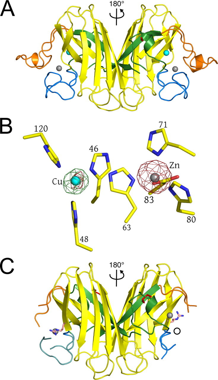
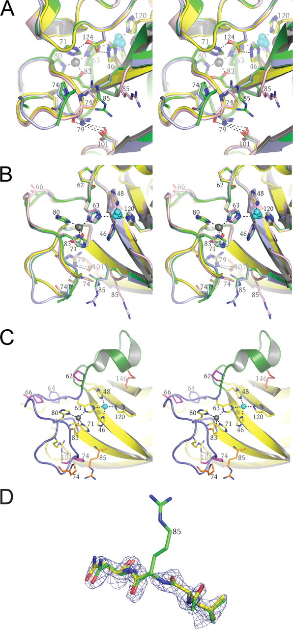
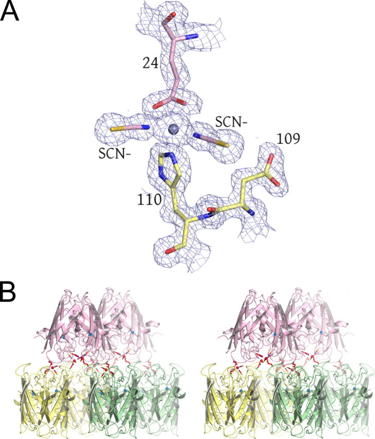
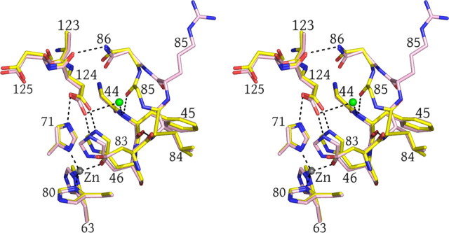
Mutations in the gene encoding human copper-zinc superoxide dismutase (SOD1) cause a dominant form of the progressive neurodegenerative disease amyotrophic lateral sclerosis. Transgenic mice expressing the human G85R SOD1 variant develop paralytic symptoms concomitant with the appearance of SOD1-enriched proteinaceous inclusions in their neural tissues. The process(es) through which misfolding or aggregation of G85R SOD1 induces motor neuron toxicity is not understood. Here we present structures of the human G85R SOD1 variant determined by single crystal x-ray diffraction. Alterations in structure of the metal-binding loop elements relative to the wild type enzyme suggest a molecular basis for the metal ion deficiency of the G85R SOD1 protein observed in the central nervous system of transgenic mice and in purified recombinant G85R SOD1. These findings support the notion that metal-deficient and/or disulfide-reduced mutant SOD1 species contribute to toxicity in SOD1-linked amyotrophic lateral sclerosis.


Figures

FIGURE 1.
FIGURE 1.
Structural comparison of human wild type and pathogenic G85R SOD1. A, wild type human SOD1. The Greek key β-barrel is shown in yellow. The electrostatic and zinc loop elements are shown in orange and blue, respectively. The disulfide loop and β-strand 8 that comprise the bulk of the dimer interface are shown in green. The disulfide bond is shown in red. Copper and zinc ions are represented as cyan and gray spheres, respectively. The relationship between the two subunits of the dimer is indicated. B, identification of copper and zinc ions in G85R SOD1 structure I, subunit A (see Table 2) using x-rays tuned to the copper and zinc absorption edges. X-rays tuned to the copper edge do not promote anomalous scattering by zinc and thereby act as the discriminator between the two metal ions. Copper does absorb slightly at the zinc edge. The green electron density is calculated from the copper edge, and the red electron density is calculated from the zinc edge. Note that the green electron density is only found around the metal in the copper site, indicating that it is copper and that there is no copper in the zinc site. The small amount of red electron density around the metal in the copper site comes from the slight anomalous scattering by copper at the zinc edge. Occupancies are estimated by comparing the thermal parameters of the metal ions and their liganding atoms. Fully occupied metal-binding sites should have thermal parameters for metal and ligand atoms that are very similar. The copper edge and zinc edge anomalous difference Fourier electron density maps are contoured at 5σ. C, human G85R SOD1 structure II, subunits E and F (see Table 2). The color coding is as in A except that the Arg85 side chains are shown as light pink and blue tubes. Zinc is bound to the copper site in the right subunit F, and the open circle indicates the position zinc would be if it were bound to the zinc site of protein as in A. The electrostatic and zinc loop elements are not shown because they are disordered in the crystal structure.
FIGURE 2.
FIGURE 2.
The G85R mutation site and the copper- and zinc-binding sites. The wild type protein is shown in yellow. A, three of the four conformations observed in the 10 unique subunits of the four crystal structures are shown in light blue, green, and pink, respectively (see text). Copper and zinc ions are represented as cyan and gray spheres, respectively. The dual hydrogen bonds formed by Asp124 to the copper ligand His46 and the zinc ligand His71 as well as the hydrogen bonding network between Pro74, Arg79, and Asp101 are shown as dotted lines. B, the image is the same as in A except rotated ∼90° around the horizontal and vertical axes in the plane of the page. The electrostatic loop has been removed for clarity. C, the location of the three proline residues of the zinc loop (see text). The disulfide loop (residues 50-62), a substructure of the zinc loop, is shown in green. The remainder of the zinc loop containing the zinc-binding ligands, residues 63-83, is shown in blue. Pro62, Pro66, and Pro74 in the zinc-bound conformation of the zinc loop are shown in magenta. The altered position of the five-membered ring of Pro74 and the Arg85 side chain are shown in orange. The hydrogen bond normally found between the carbonyl oxygen of Pro74 and the guanidinium group of Arg79 is disrupted. D, structural state four (see text) found in G85R subunits C, E, and F (see Table 2). The absence of electron density around the α carbon and the Arg85 side chain indicates that this residue is conformationally mobile, sampling many positions. This movement is correlated with both zinc deficiency in the zinc site and disorder of the zinc and electrostatic loop elements.
FIGURE 3.
FIGURE 3.
Surface zinc-binding site observed in structures IV and V. A, the bridging zinc ion between two G85R SOD1 dimers is shown as a gray sphere. Residues coming from a subunit of one dimer are shown in yellow, and those coming from another dimer are shown in pink. The thiocyanate anions are labeled. The SIGMAA electron density is contoured at 1.5σ. B, the bridging zinc ions link each SOD1 dimer to other dimers through a 2-fold rotation and a translation of one subunit. The bridging zinc ion and its ligands are shown in red. These interactions are propagated throughout the entire crystal along the b axis. The zinc ion in the zinc-binding site is shown as a blue sphere where it caps the helix dipole at the C terminus of the short α-helix in the electrostatic loop.
FIGURE 4.
FIGURE 4.
A novel water molecule gains access to the active site area in the three subunits with a displaced 85-86 peptide bond (see also Table 2 and the text). The wild type enzyme is shown in yellow, and the G85R SOD1 mutant is shown in pink. The water molecule gaining access to zinc site in the G85R structure is shown as a green sphere.

References

    1. McCord, J. M., and Fridovich, I. (1969) J. Biol. Chem. 244 6049-6055 - PubMed
    1. Deisseroth, A., and Dounce, A. L. (1970) Physiol. Rev. 50 319-375 - PubMed
    1. Little, C., and O'Brien, P. J. (1968) Biochem. Biophys. Res. Commun. 31 145-150 - PubMed
    1. Marklund, S. L. (1984) J. Clin. Investig. 74 1398-1403 - PMC - PubMed
    1. Pardo, C. A., Xu, Z., Borchelt, D. R., Price, D. L., Sisodia, S. S., and Cleveland, D. W. (1995) Proc. Natl. Acad. Sci. U. S. A. 92 954-958 - PMC - PubMed

Publication types

MeSH terms

Associated data