. 2008 Jun;28(11):3639-51.
doi: 10.1128/MCB.01828-07. Epub 2008 Mar 31.

Molecular characterization of the role of the Schizosaccharomyces pombe nip1+/ctp1+ gene in DNA double-strand break repair in association with the Mre11-Rad50-Nbs1 complex

Affiliations

Molecular characterization of the role of the Schizosaccharomyces pombe nip1+/ctp1+ gene in DNA double-strand break repair in association with the Mre11-Rad50-Nbs1 complex

Yufuko Akamatsu et al. Mol Cell Biol. 2008 Jun.

Abstract

The Schizosaccharomyces pombe nip1(+)/ctp1(+) gene was previously identified as an slr (synthetically lethal with rad2) mutant. Epistasis analysis indicated that Nip1/Ctp1 functions in Rhp51-dependent recombinational repair, together with the Rad32 (spMre11)-Rad50-Nbs1 complex, which plays important roles in the early steps of DNA double-strand break repair. Nip1/Ctp1 was phosphorylated in asynchronous, exponentially growing cells and further phosphorylated in response to bleomycin treatment. Overproduction of Nip1/Ctp1 suppressed the DNA repair defect of an nbs1-s10 mutant, which carries a mutation in the FHA phosphopeptide-binding domain of Nbs1, but not of an nbs1 null mutant. Meiotic DNA double-strand breaks accumulated in the nip1/ctp1 mutant. The DNA repair phenotypes and epistasis relationships of nip1/ctp1 are very similar to those of the Saccharomyces cerevisiae sae2/com1 mutant, suggesting that Nip1/Ctp1 is a functional homologue of Sae2/Com1, although the sequence similarity between the proteins is limited to the C-terminal region containing the RHR motif. We found that the RxxL and CxxC motifs are conserved in Schizosaccharomyces species and in vertebrate CtIP, originally identified as a cofactor of the transcriptional corepressor CtBP. However, these two motifs are not found in other fungi, including Saccharomyces and Aspergillus species. We propose that Nip1/Ctp1 is a functional counterpart of Sae2/Com1 and CtIP.


Figures
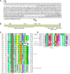
FIG. 1.
FIG. 1.
Nip1 protein motifs. (A) Structure of the nip1 gene. The isolated cDNA contains nucleotides −429 to +958. The ORF of nip1 consists of two exons separated by one intron. Three serine residues and one threonine residue in consensus phosphorylation site sequences for ATM/ATR and CDK are indicated by circles. Underlined sequences are highly conserved sequences; one contains the RxxL and CxxC motifs, and the other contains the Sae2 RHR motif. (B) Schematic presentation of the S. pombe Nip1 protein. The serine at position 35 and the threonine at position 175 are potential phosphorylation sites for ATM/ATR kinases, and the two serines at positions 114 and 165 are potential CDK phosphorylation sites. The N-terminal region (22 to 160) is predicted to form a coiled-coil structure by the COILS program (40). (C) Sequence alignment of the region containing the Sae2 RHR motif. The consensus sequence is (G/S/T)RHR. The Sae2 motif is found in many eukaryotes, including fungi. (D) Sequence alignment of the region containing the RxxL (D-box) and CxxC motifs of two fission yeast, S. pombe and Schizosaccharomyces japonicus, five vertebrates, two higher plants, a nematode, and two protists. Abbreviations for the species whose sequences are shown in panels C and D and accession numbers for the Nip1 homologues are as follows: Spo, S. pombe; Sja, S. japonicus; nig, Aspergillus niger (CAK45154); Aor, Aspergillus oryzae (BAE61104); Afu, Aspergillus fumigatus (XP_750508); Acl, Aspergillus clavatus (XP_001269402); Ate, Aspergillus terreus (XP_001209515); nid, Aspergillus nidulans (XP_662716); Aca, Ajellomyces capsulatus (XP_001545011); Cim, Coccidioides immitis RS (XP_001248767); Bfu, Botryotinia fuckeliana (XP_001552631); Ssc, Sclerotinia sclerotioru (EDO02392); Pno, Phaeosphaeria nodorum SN15 (EAT90897); Ncr, Neurospora crassa (XP_957865); glo, Chaetomium globosum (EAQ85759); Yli, Yarrowia lipolytica (XP_502193); Cne, Cryptococ-cus neoformans (XP_572699); Cci, Coprinopsis cinerea (EAU86571); Uma, Ustilago maydis (XP_758978); Hsa, Homo sapiens (AAC14371); Mmu, Mus musculus (house mouse, NP_001074692); Gga, Gallus gallus (XP_419158); Xla, Xenopus laevis (African clawed frog, NP_001085825); Dre, Danio rerio (zebra fish, NP_001012518); Ath, Arabidopsis thaliana (Q9ZRT1); Osa, Oryza sativa (EAZ01661); Ddi, Dictyostelium discoideum (XP_635231); Cel, C. elegans (NP_499398); Cpa, Cryptosporidium parvum (XP_628681); Sce, S. cerevisiae (CAA96887); Sca, Saccharomyces cariocanus (ABI48902); Spa, Saccharomyces pastorianus (ABI48905); Vpo, Vanderwaltozyma polyspora (EDO17800); Dha, Debaryomyces hansenii (CAG86377); Ago, Ashbya gossypii (NP_984048); Kla, Kluyveromyces lactis (CAG98790); gla, Candida glabrata (CAG62002).
FIG. 2.
FIG. 2.
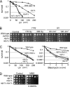
Genetic interactions of Nip1 with Rhp51 and the MRN complex. (A) Killing curve for UV irradiation suggests that Nip1 functions in the Rhp51-dependent repair pathway. The strains used were SP129 (wild type, crosses), YA1097 (nip1Δ, closed circles), YA1177 (rhp51Δ, squares), and YA1268 (nip1Δ rhp51Δ, open circles). (B) The rad32Δ (YA1077), nbs1Δ (SPN341), and nip1Δ (YA1097) single mutants showed very similar sensitivities to γ and UV irradiation. SP129 was used as the wild-type control. (C and D) A nip1Δ nbs1Δ double mutant showed sensitivities to UV, bleomycin, and MMS that were similar to those of the single mutants. The strains used were SP129 (wild type, crosses), YA1097 (nip1Δ, open circles), YA1184 (nbs1Δ, squares), and YA1233 (nip1Δ nbs1Δ, closed circles).
FIG. 3.
FIG. 3.
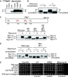
Isolation and characterization of the nbs1-s10 allele. (A) Two plasmids restored normal MMS resistance to an slr10 mutant (no. 16). Two mutant no. 16 transformants, transformed by clone 2-36 (nbs1+) and clone 7 (nip1+), exhibit a sensitivity to MMS (0.004%) similar to that of the wild-type strain (SP185). (B) Sequence alignment of the FHA domain of human (h), mouse (m), and S. pombe (sp) Nbs1, and S. cerevisiae (sc) Xrs2. The arrowhead indicates the conserved Gly103 residue that is mutated to Asp in the slr10 mutant. (C) Expression of wild-type Nbs1 and Nbs1-s10 proteins detected by Western blotting. Wild-type (SP129), nbs1-s10 (YA1128), and nbs1Δ (YA1184) cells were analyzed. (D) The sensitivity to UV and bleomycin of the nbs1-s10 mutant was intermediate between those of the control and nbs1Δ mutant strains. The strains used for the spot tests were the same as in panel C. (E) nbs1+, but not nbs1-s10, fully complemented the DNA damage sensitivity of the nbs1-s10 mutant. Wild-type (WT, SP129) and nbs1-s10 (YA1128) cells were transformed with the pSP102 vector alone, pSP102 carrying nbs1-s10, or pSP102 carrying nbs1+, and the sensitivity of the transformants to HU, UV, bleomycin, and MMS was analyzed. (F) The DNA damage sensitivity of the nbs1-s10 mutant, but not of the nbs1Δ mutant, was suppressed by nip1+ expressed from a multicopy plasmid, pSP102. Wild-type (SP129), nbs1-s10 (YA1128), and nbs1Δ (YA1184) cells were transformed with the pSP102 vector, pSP102 carrying nip1+, or pSP102 carrying nbs1+, and the sensitivity of the transformants to HU, UV, bleomycin, and MMS was analyzed.
FIG. 4.
FIG. 4.
Nip1 is phosphorylated under normal growth conditions and becomes more extensively phosphorylated in response to treatments that cause DNA damage. (A) Nip1 is modified upon DNA damage. Cell extracts from an untagged Nip1 strain (YA119) and a Nip1-FLAG strain (YST153) were analyzed by Western blotting with an anti-FLAG antibody. Under normal conditions, Nip1 migrates to the Nip1-F position. Upon DNA damage, modified Nip1 protein migrates to the position indicated by Nip1-S. non, nontreated; bleo, bleomycin. (B) Phosphatase treatment reveals that the Nip1 modification is a double phosphorylation. Immunocomplexes from the Nip1-FLAG strain were treated with lambda protein phosphatase as described in Materials and Methods. S and F in panels A and B indicate Nip1-S and Nip1-F, respectively. (C) Nip1 mutants with mutations at four possible phosphorylation sites are still phosphorylated upon bleomycin treatment. (Top) Wild-type Nip1 protein has four serine/threonine residues that are potential phosphorylation sites. Combinations of these residues, S35-Q36, S114-P115, S165-P166, and T175-Q176, were mutated to alanines. (Bottom) Three Nip1-FLAG mutants were constructed, an S35A-T175A double mutant named SQ/TQ (YST442), an S114A-S165A double mutant named SP1/SP2 (YST445), and an S114A-S165A-SP1/SP2 quadruple mutant named SQ/TQ/SP1/SP2 (YST447). A wild-type strain (YST438) and the mutant strains were incubated in the presence (+) or absence (−) of bleomycin, and cell extracts were analyzed by Western blotting with the anti-FLAG antibody. The SP1/SP2 and SQ/TQ/SP1/SP2 mutant proteins migrated to the position corresponding to that of fully dephosphorylated Nip1 (Nip1-FF) but were modified upon treatment with bleomycin and migrated to the Nip1-F position. aa, amino acids. (D) Phosphatase treatment reveals that the modification of the SP1/SP2 mutant protein is phosphorylation. Immunocomplexes from the SP1/SP2-FLAG strain (YST445) were treated with lambda protein phosphatase as described in Materials and Methods. (E) The SQ/TQ (YST442), SP1/SP2 (YST445), and SQ/TQ/SP1/SP2 (YST447) mutants do not show a detectable DNA repair defect.
FIG. 5.
FIG. 5.
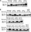
Genetic requirements of Nip1 phosphorylation. (A) Rad32 and Nbs1, but not Rhp51, are required for the DNA damage-induced phosphorylation of Nip1. (B) Tel1 and Rad3 are required for the DNA damage-induced phosphorylation of Nip1, whereas other downstream checkpoint factors are not essential for phosphorylation.
FIG. 6.
FIG. 6.
Meiotic DSBs accumulate in nip1Δ mutant cells. pat1-114 haploid cells with various mutations were induced to synchronously undergo meiosis and collected at the indicated time points, and chromosomal DNA was analyzed by PFGE. wt, wild type; Chr., chromosome.

References

    1. Akamatsu, Y., D. Dziadkowiec, M. Ikeguchi, H. Shinagawa, and H. Iwasaki. 2003. Two different Swi5-containing protein complexes are involved in mating-type switching and recombination repair in fission yeast. Proc. Natl. Acad. Sci. USA 10015770-15775. - PMC - PubMed
    1. Alani, E., R. Padmore, and N. Kleckner. 1990. Analysis of wild-type and rad50 mutants of yeast suggests an intimate relationship between meiotic chromosome synapsis and recombination. Cell 61419-436. - PubMed
    1. Amador, V., S. Ge, P. G. Santamaria, D. Guardavaccaro, and M. Pagano. 2007. APC/C(Cdc20) controls the ubiquitin-mediated degradation of p21 in prometaphase. Mol. Cell 27462-473. - PMC - PubMed
    1. Arora, C., K. Kee, S. Maleki, and S. Keeney. 2004. Antiviral protein Ski8 is a direct partner of Spo11 in meiotic DNA break formation, independent of its cytoplasmic role in RNA metabolism. Mol. Cell 13549-559. - PubMed
    1. Baroni, E., V. Viscardi, H. Cartagena-Lirola, G. Lucchini, and M. P. Longhese. 2004. The functions of budding yeast Sae2 in the DNA damage response require Mec1- and Tel1-dependent phosphorylation. Mol. Cell. Biol. 244151-4165. - PMC - PubMed

Publication types

MeSH terms