. 2008 Jun;28(11):3830-49.
doi: 10.1128/MCB.01217-07. Epub 2008 Mar 31.

Progesterone induction of the 11beta-hydroxysteroid dehydrogenase type 2 promoter in breast cancer cells involves coordinated recruitment of STAT5A and progesterone receptor to a distal enhancer and polymerase tracking

Affiliations

Progesterone induction of the 11beta-hydroxysteroid dehydrogenase type 2 promoter in breast cancer cells involves coordinated recruitment of STAT5A and progesterone receptor to a distal enhancer and polymerase tracking

Alicia Subtil-Rodríguez et al. Mol Cell Biol. 2008 Jun.

Abstract

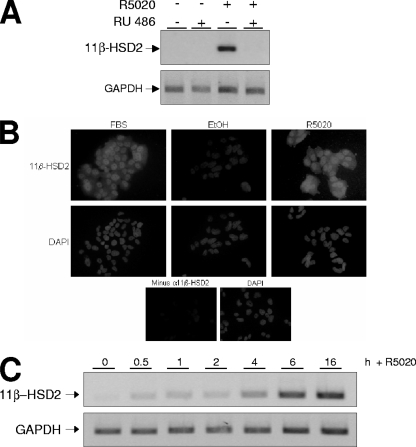
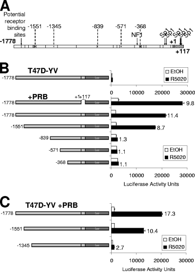
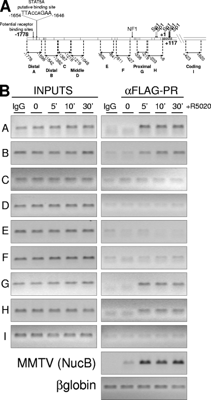
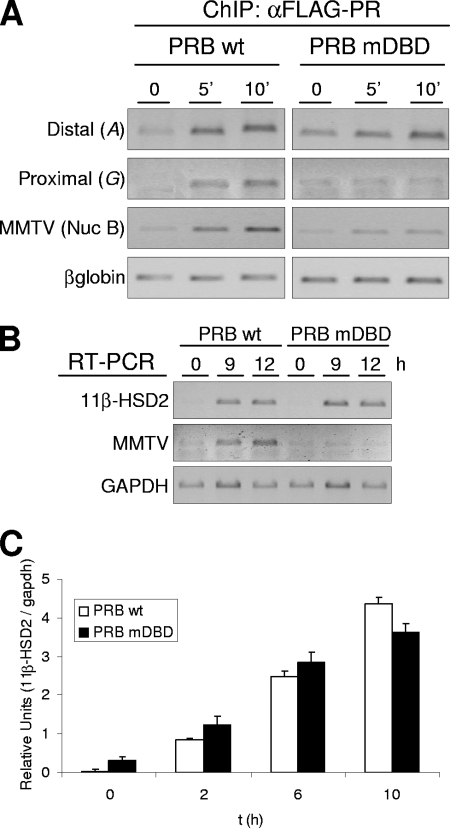
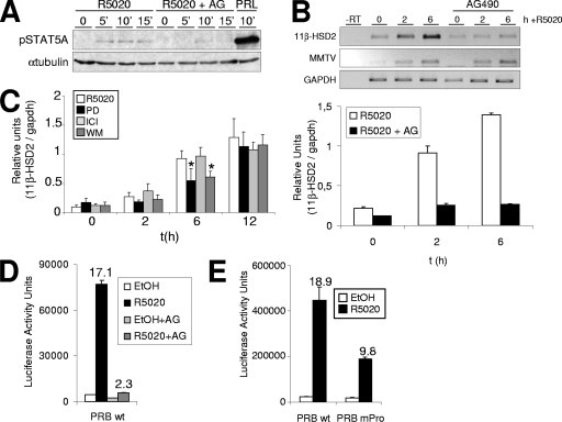
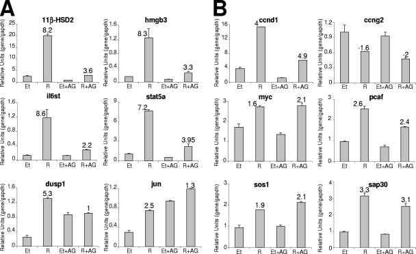
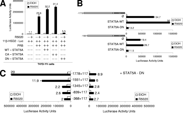
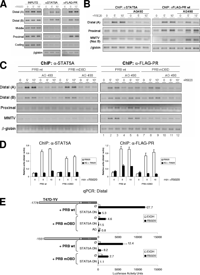
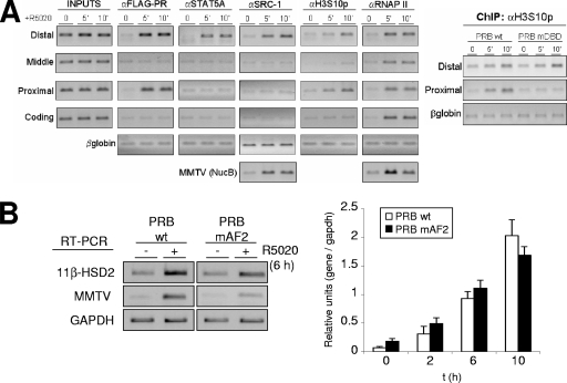
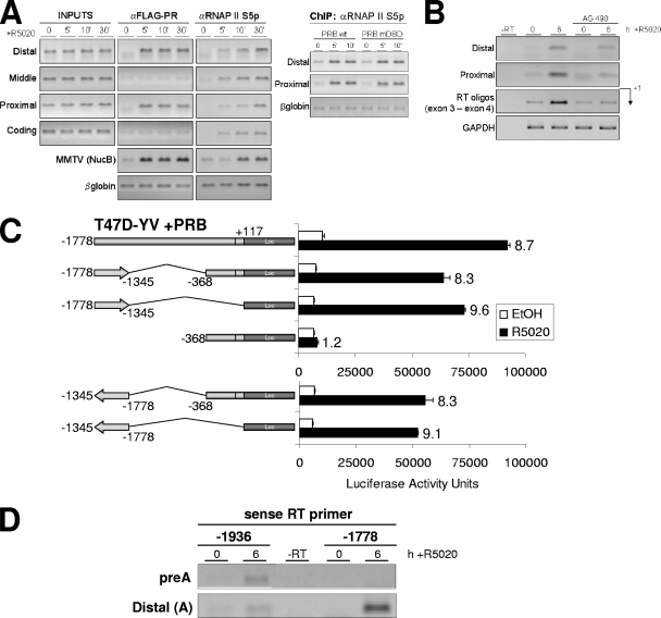
Steroid hormone receptors regulate gene expression, interacting with target DNA sequences but also activating cytoplasmic signaling pathways. Using the human 11beta-hydroxysteroid dehydrogenase type 2 (11beta-HSD2) gene as a model, we have investigated the contributions of both effects on a human progesterone-responsive promoter in breast cancer cells. Chromatin immunoprecipitation has identified two different mechanisms of hormone-induced progesterone receptor (PR) recruitment to the 11beta-HSD2 promoter: (i) direct PR binding to DNA at the proximal promoter, abrogated when PR contains a mutated DNA binding domain (DBD), and (ii) STAT5A (signal transducer and activator of transcription 5A)-mediated recruitment of PR to an upstream distal region, impaired by AG490, a JAK/STAT pathway inhibitor. The JAK/STAT inhibitor, as well as expression of dominant-negative STAT5A, impairs hormone induction of 11beta-HSD2. On the other hand, the DBD-mutated PR fully supports 11beta-HSD2 expression. These results, along with data from a deletion analysis, indicate that the distal region is crucial for hormone regulation of 11beta-HSD2. We show active RNA polymerase II tracking from the distal region upon PR and STAT5A binding, concomitant with synthesis of noncoding, hormone-dependent RNAs, suggesting that this region works as a hormone-dependent transcriptional enhancer. In conclusion, coordination of PR transcriptional effects and cytoplasmic signaling activation, in particular the JAK/STAT pathway, are critical in regulating progestin-induced endogenous 11beta-HSD2 gene expression in breast cancer cells. This is not unique to this promoter, as AG490 also alters the expression of other progesterone-regulated genes.


Figures

FIG. 1.
FIG. 1.
11β-HSD2 promoter expression in T47D cells is induced by progestin and depends on the classical PR. (A) T47D cells were cultured in phenol red-free medium under serum-free conditions for 48 h before the addition of R5020 (10 nM) and/or RU (1 μM) or vehicle for 16 h. Cells were harvested, and total RNA was extracted. The 11β-HSD2 mRNA expression was analyzed by RT-PCR with specific primers. GAPDH cDNA-specific primers were used as a control. PCR products were run on a 1.2% agarose gel and visualized with ethidium bromide. (B) Hormone-induced accumulation of 11β-HSD2 enzyme detected by immunofluorescence. T47D cells were cultured over coverslips in 10% FBS rich medium or in serum-free, steroid-deprived medium for 48 h before the addition of R5020 (10 nM) or ethanol (EtOH) for 24 h. Then, the cells were fixed, impermeabilized, and incubated with anti-11β-HSD2 antibody, followed by incubation with biotinylated secondary antibody and then streptavidin-conjugated Alexa 488 and, finally, DAPI, to stain nucleic acids (as described in Materials and Methods). (Lower panels) For a negative control, T47D cells cultured in rich medium were analyzed as previously described in the absence of the primary antibody. (C) T47D cells, cultured as described for panel A, were untreated (0) or treated with R5020 (10 nM) for the times indicated. 11β-HSD2 expression was analyzed as described for panel A, and GAPDH expression was used as a control.
FIG. 2.
FIG. 2.
An 11β-HSD2 promoter region upstream of nucleotide −1345 is necessary for progesterone-induced activation. (A) Human 11β-HSD2 promoter region structure. Arrows indicate the positions of NF1 and Sp1 binding sites. The vertical bars along the 11β-HSD2 promoter region indicate the positions of potential progesterone receptor elements predicted by computational analysis with ConSite and Transfac software. The deletion endpoints used in the deletion analysis are indicated. (B, C) Analysis of promoter deletion constructs driving Luc reporter expression. Indicated constructs (−1778, −1551, −839, −571, and −368 in panel B; −1778, −1551, and −1345 in panel C) were transfected together with a PRB expression vector into T47D-YV cells. After transfection, cells were serum starved for 48 h and then treated with ethanol (EtOH) or R5020 (10 nM) for 16 h and Luc activity was measured. For normalization, equal amounts of extracted protein of each sample were used. Data of a representative experiment performed in duplicate, with each sample analyzed in duplicate, are expressed as mean numbers of Luc arbitrary units, and error bars represent the data ranges. Induction in response to hormone compared to the level for ethanol is shown for each construct.
FIG. 3.
FIG. 3.
PR binds to two different regions of the 11β-HSD2 promoter after hormone activation. (A) Schematic representation of the 11β-HSD2 promoter region with the locations of amplicons for ChIP analysis as well as the location and sequence of the potential STAT5A binding site as predicted by ConSite and Transfac software. (B) TYML cells (T47D-YV cell line carrying an integrated copy of the progesterone reporter MMTV-Luc), expressing FLAG-tagged WT PRB, were cultured as described for Fig. 1A, untreated (0) or treated with R5020 (10 nM) for 5, 10, or 30 min, harvested, and used for ChIP experiments with anti-FLAG antibody. The precipitated DNA fragments were subjected to PCR with primers for the indicated 11β-HSD2 promoter regions or the β-globin gene as a control. The MMTV nucleosome B region was also amplified to detect PR recruitment as a control for the experiment. Amplification of input DNA (representing 1% of immunoprecipitated material) is shown for comparison. PCR products were run in a 1.2% agarose gel and visualized with ethidium bromide.
FIG. 4.
FIG. 4.
Effect of PR mutations at the DBD on progesterone-induced 11β-HSD2 promoter activity. (A) TYML cells expressing FLAG-tagged WT PRB or mutant DBD PRB (PRB-mDBD), cultured as described for Fig. 1A, were untreated (0) or treated with R5020 (10 nM) for 5 or 10 min, harvested, and used for ChIP experiments with anti-FLAG tag (αFLAG) antibody. The precipitated DNA fragments were subjected to PCR analysis with specific primers corresponding to the distal or proximal 11β-HSD2 promoter region, with MMTV nucleosome B and the β-globin gene as a control. (B) TYML cells expressing WT PRB or PRB-mDBD, cultured as described for Fig. 1A, were untreated (0) or treated with R5020 (10 nM) for 9 or 12 h. Cells were harvested, and total RNA was extracted. The 11β-HSD2 and MMTV-Luc mRNA expression levels were analyzed by RT-PCR with specific primers. GAPDH cDNA-specific primers were used as a control. PCR products were run on a 1.2% agarose gel and visualized with ethidium bromide. (C) TYML cells expressing WT PRB or PRB-mDBD, cultured as described for Fig. 1A, were untreated (0) or treated with R5020 (10 nM) for 2, 6, or 10 h. Cells were then harvested, and total RNA was extracted. 11β-HSD2 mRNA expression was analyzed by RT and real-time PCR with specific primers. GAPDH expression was measured for normalization. Data of a representative experiment performed in duplicate, with each sample analyzed in duplicate, are expressed as mean numbers of 11β-HSD2/GAPDH relative units, and error bars represent the data ranges.
FIG. 5.
FIG. 5.
JAK/STAT pathway activation is involved in the hormonal induction of 11β-HSD2 gene expression. (A) T47D cells were cultured in phenol red-free medium under serum-free conditions for 72 h prior to the addition of 10 nM R5020 for 5, 10, or 15 min or 500 ng/ml prolactin (PRL) for 10 min. When indicated, 1 h before hormonal induction, cells were treated with 50 μM AG. Cell lysates were analyzed by Western blotting with antibodies against phosphorylated STAT5A (Tyr694/699) and tubulin. (B) T47D cells, cultured as described for Fig. 1A, were treated with R5020 (10 nM) for 0, 2, or 6 h. When indicated, AG (50 μM) was added 1 h before hormone addition. Cells were harvested, and total RNA was extracted. (Upper panel) 11β-HSD2 and MMTV-Luc mRNA expression levels were analyzed by RT-PCR with specific primers. GAPDH cDNA-specific primers were used as a control. PCR products were run on a 1.2% agarose gel and visualized with ethidium bromide. (Lower panel) 11β-HSD2 mRNA expression was analyzed by RT and real-time PCR with specific primers. GAPDH cDNA-specific primers were used as a control. The values represent the means and ranges of a representative experiment performed in duplicate, with each sample analyzed in duplicate, expressed as numbers of 11β-HSD2/GAPDH relative units. (C) Involvement of MAPK and PI3K pathways in hormone activation of 11β-HSD2. T47D cells, cultured as described for Fig. 1A, were untreated (0) or treated with R5020 (10 nM) for 2, 6, or 12 h. When indicated, PD (50 μM), WM (0.1 μM), or ICI (10 μM) was added 1 h before hormone induction. Cells were harvested, total RNA was prepared, and cDNA was generated by RT. 11β-HSD2 expression was analyzed by real-time PCR with specific primers. To normalize the data, GAPDH expression was used as a control. The values represent the means and standard deviations of two experiments performed in duplicate, expressed as numbers of 11β-HSD2/GAPDH relative units. Asterisks denote significant differences (P < 0.05) between treated (PD or WM) and untreated (only R5020) data sets, as analyzed by Student's t test. (D) T47D-YV cells cotransfected with WT pSG5-PRB and reporter 11β-HSD2-Luc vectors were treated with R5020 (10 nM) for 16 h, and Luc activity was determined. When indicated, cells were treated with AG (50 μM) 1 h before hormone addition. For normalization, equal amounts of cellular extract of each sample were used. The values represent the mean numbers of arbitrary Luc units and ranges of a representative experiment performed in duplicate. EtOH, ethanol. (E) Expression of 11β-HSD2 reporter construct in the presence of a Pro cluster PR mutant. T47D-YV cells cotransfected with WT pSG5-PRB or a Pro cluster mutant and reporter 11β-HSD2-Luc vectors were treated with R5020 (10 nM) for 16 h, and Luc activity was determined. For normalization, equal amounts of cellular extract of each sample were used. The values represent the mean numbers of arbitrary Luc units and ranges of a representative experiment performed in duplicate.
FIG. 6.
FIG. 6.
Involvement of JAK/STAT pathway activation in progestin-induced gene expression. T47D cells, cultured as described for Fig. 1A, were treated for 6 h with R5020 (10 nM) or ethanol (Et), and when indicated, AG was added 1 h before hormone. Cells were harvested, and total RNA was extracted. Expression of the genes indicated was analyzed by RT and real-time PCR with specific primers. GAPDH cDNA-specific primers were used as a control. The values represent the means and ranges of a representative experiment performed in duplicate, expressed as numbers of specific gene/GAPDH relative units. Inductions in response to hormone are indicated.
FIG. 7.
FIG. 7.
Expression of DN STAT5A affects the hormone activation of the 11β-HSD2 promoter. (A) T47D-YV cells were cotransfected when indicated with 1 μg of 11β-HSD2-Luc (−1778/+117) reporter vector, 1 μg of pSG5-PRB, and 1 μg of WT STAT5A, CA STAT5A, or DN STAT5A. Afterwards, cells were treated with ethanol (EtOH) or R5020 (10 nM) for 16 h and Luc activity was measured. For normalization, equal amounts of cellular extract of each sample were used. The values represent the mean numbers of arbitrary Luc units and ranges of a representative experiment performed in duplicate. Induction in response to hormone is shown for each construct. (B, C) T47D-YV (B) or T47D (C) cells cotransfected with 1 μg of the indicated 11β-HSD2 promoter constructs, 1 μg of pSG5-PRB, and 1 μg of plasmid expressing WT STAT5A or DN STAT5A were treated with ethanol or R5020 (10 nM) for 16 h, and Luc activity was measured. Induction in response to hormone is shown for each construct. The values represent the means and ranges of a representative experiment performed in duplicate.
FIG. 8.
FIG. 8.
STAT5A and PR recruitment to the distal region depends on the JAK/STAT pathway. (A) TYML cells expressing FLAG-tagged WT PRB were cultured as described for Fig. 1A, untreated (0) or treated with R5020 (10 nM) for 5 or 10 min, harvested, and used for ChIP experiments using anti-STAT5A (αSTAT5A; middle panel) or anti-FLAG tag (right panel) antibodies. The precipitated DNA fragments were subjected to PCR analysis with primers corresponding to the indicated 11β-HSD2 promoter regions and the β-globin gene. Input material (1%) is shown for comparison. PCR products were run in a 1.2% agarose gel and visualized with ethidium bromide. (B) TYML cells expressing FLAG-tagged WT PRB, cultured as described for Fig. 1A, were untreated (0) or treated with R5020 (10 nM) for 5 or 10 min, harvested, and used for ChIP experiments with anti-STAT5A or anti-FLAG-tag antibodies. When indicated, cells were treated with AG (50 μM) 1 h before hormone addition. The precipitated DNA fragments were subjected to PCR analysis with specific primers corresponding to the distal and proximal 11β-HSD2 promoter regions or MMTV nucleosome B and the β-globin gene as a control. PCR products were run in a 1.2% agarose gel and visualized with ethidium bromide. (C) ChIP experiment as in panel B from TYML cells expressing FLAG-tagged WT PRB or mutant DBD PRB (PRB-mDBD), combined with AG (50 μM). Here, the average chromatin fragment size was 200 bp (see Materials and Methods). (D) Real-time PCR quantification of ChIP samples from panel C for distal amplicon A, performed in duplicate and normalized to actin gene amplification levels. The values represent the means and data ranges. (E) T47D-YV cells cotransfected with 1 μg of the 11β-HSD2 −1778 or −1551 promoter construct, 1 μg of WT or PRB-mDBD, and 1 μg of DN STAT5A when indicated were treated with ethanol (EtOH) or R5020 (10 nM) for 16 h, and Luc activity was measured. Where indicated, 1 h of AG pretreatment was performed. Induction in response to hormone is shown for each construct. The values represent the means and ranges of a representative experiment performed in duplicate.
FIG. 9.
FIG. 9.
Coactivator recruitment and histone modifications at the 11β-HSD2 promoter. (A) TYML cells expressing FLAG-tagged WT PRB, cultured as described for Fig. 1A, were untreated (0) or treated with R5020 (10 nM) for 5 or 10 min, harvested, and used for ChIP experiments with anti-FLAG tag (αFLAG), STAT5A, SRC-1, H3S10p, and RNAP II antibodies. The precipitated DNA fragments were subjected to PCR analysis with specific primers corresponding to the indicated 11β-HSD2 promoter regions or MMTV nucleosome B and the β-globin gene as a control. For the right panel, ChIP with H3S10p antibody was performed on chromatin extracted from TYML cells expressing FLAG-tagged WT PRB or PRB-mDBD, untreated or treated with R5020. (B) TYML cells expressing WT PRB or PRB-mAF2, cultured as described for Fig. 1A, were untreated (0) or treated with R5020 (10 nM) for 2, 6, or 10 h. Cells were harvested, and total RNA was extracted. (Left) 11β-HSD2 and MMTV-Luc mRNA expression levels were analyzed by RT-PCR with specific primers. GAPDH cDNA-specific primers were used as a control. PCR products were run on a 1.2% agarose gel and visualized with ethidium bromide. (Right) 11β-HSD2 mRNA expression was analyzed by RT and real-time PCR with specific primers. GAPDH cDNA-specific primers were used as a control. The values represent the means and ranges of a representative experiment performed in duplicate, expressed as numbers of 11β-HSD2/GAPDH relative units.
FIG. 10.
FIG. 10.
The distal region works as a hormone-responsive RNAP II entry site from where RNA synthesis occurs and enhances 11β-HSD2 expression. (A) TYML cells expressing WT FLAG-tagged PRB, cultured as described for Fig. 1A, were untreated (0) or treated with R5020 (10 nM) for 5 or 10 min, harvested, and used for ChIP experiments with anti-FLAG tag (αFLAG) and phospho(Ser5)-RNAP II (RNAP II S5p) antibodies. The precipitated DNA fragments were subjected to PCR analysis with specific primers corresponding to the indicated 11β-HSD2 promoter regions or MMTV nucleosome B and the β-globin gene as a control. On the right panel, anti-phospho-RNAP II ChIP was performed on chromatin extracted from TYML cells expressing FLAG-tagged WT PRB or PRB-mDBD, untreated or treated with R5020. (B) TYML cells expressing FLAG-tagged WT PRB, cultured as described for Fig. 1A, were treated with ethanol or R5020 (10 nM) for 6 h. When indicated, cells were pretreated with AG (50 μM). Cells were harvested, total RNA was prepared, and cDNA was generated by RT using oligo(dT). RNA synthesis was analyzed using primers that specifically amplified the 11β-HSD2 promoter distal region and the proximal region and primers located in exon 3 and exon 4 (RT oligos). GAPDH-specific primers were used as a control. (C) T47D-YV cells cotransfected with 1.5 μg of WT pSG5-PRB expression vector, 1.5 μg of the indicated 11β-HSD2 deletion constructs was treated with ethanol (EtOH) or R5020 (10 nM) for 16 h, and Luc activity was measured. For normalization, equal amounts of cellular extract of each sample were used. The values represent the mean numbers of arbitrary Luc units and ranges of a representative experiment performed in duplicate. Inductions in response to hormone are indicated. (D) TYML cells expressing FLAG-tagged WT PRB, cultured as described for Fig. 1A, were treated with EtOH or R5020 (10 nM) for 6 h. Then, cells were harvested, total RNA was prepared, and cDNA was generated by RT using gene-specific sense primers at positions −1936 and −1778 upstream of the TSS. The presence of antisense RNA upstream of the TSS was analyzed using PCR primers at positions −1936/−1779 (preA) and −1778/−1596 (distal A) that specifically amplified the promoter region.

Similar articles

Cited by

References

    1. Agarwal, A. K., and P. C. White. 1996. Analysis of the promoter of the NAD+ dependent 11 beta-hydroxysteroid dehydrogenase (HSD11K) gene in JEG-3 human choriocarcinoma cells. Mol. Cell. Endocrinol. 12193-99. - PubMed
    1. Albiston, A. L., V. R. Obeyesekere, R. E. Smith, and Z. S. Krozowski. 1994. Cloning and tissue distribution of the human 11 beta-hydroxysteroid dehydrogenase type 2 enzyme. Mol. Cell. Endocrinol. 105R11-R17. - PubMed
    1. Alikhani-Koopaei, R., F. Fouladkou, F. J. Frey, and B. M. Frey. 2004. Epigenetic regulation of 11 beta-hydroxysteroid dehydrogenase type 2 expression. J. Clin. Investig. 1141146-1157. - PMC - PubMed
    1. Ariyoshi, K., T. Nosaka, K. Yamada, M. Onishi, Y. Oka, A. Miyajima, and T. Kitamura. 2000. Constitutive activation of STAT5 by a point mutation in the SH2 domain. J. Biol. Chem. 27524407-24413. - PubMed
    1. Ballaré, C., M. Uhrig, T. Bechtold, E. Sancho, M. Di Domenico, A. Migliaccio, F. Auricchio, and M. Beato. 2003. Two domains of the progesterone receptor interact with the estrogen receptor and are required for progesterone activation of the c-Src/Erk pathway in mammalian cells. Mol. Cell. Biol. 231994-2008. - PMC - PubMed

Publication types

MeSH terms