. 2008 Apr 14;205(4):777-83.
doi: 10.1084/jem.20072513. Epub 2008 Mar 31.

CXCR4 is required for the quiescence of primitive hematopoietic cells

Affiliations

CXCR4 is required for the quiescence of primitive hematopoietic cells

Yuchun Nie et al. J Exp Med. .

Abstract

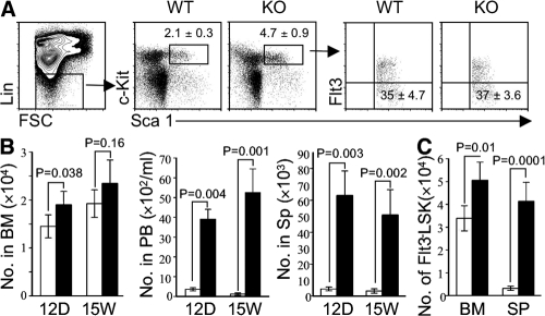
The quiescence of hematopoietic stem cells (HSCs) is critical for preserving a lifelong steady pool of HSCs to sustain the highly regenerative hematopoietic system. It is thought that specialized niches in which HSCs reside control the balance between HSC quiescence and self-renewal, yet little is known about the extrinsic signals provided by the niche and how these niche signals regulate such a balance. We report that CXCL12 produced by bone marrow (BM) stromal cells is not only the major chemoattractant for HSCs but also a regulatory factor that controls the quiescence of primitive hematopoietic cells. Addition of CXCL12 into the culture inhibits entry of primitive hematopoietic cells into the cell cycle, and inactivation of its receptor CXCR4 in HSCs causes excessive HSC proliferation. Notably, the hyperproliferative Cxcr4(-/-) HSCs are able to maintain a stable stem cell compartment and sustain hematopoiesis. Thus, we propose that CXCR4/CXCL12 signaling is essential to confine HSCs in the proper niche and controls their proliferation.


Figures

Figure 1.
Figure 1.
Cxcr4−/− HSCs are retained in the BM. (A) Dot plots represent the Flt3LSK population in the BM of Cxcr4−/− (KO) and control mice (WT) at day 12 after tamoxifen treatment. The percentages of gated populations of Flt3LSK cells ± the SD are shown. (B) Absolute numbers of Flt3LSK cells in the femur (BM), peripheral blood (PB), and spleen (Sp) of Cxcr4−/− and control mice at day 12 and week 15 after tamoxifen treatment. Values are the mean ± the SD (n = 6). (C) Absolute numbers of Flt3LSK cells in the femur and tibia (BM) and spleen (SP) 32 wk after tamoxifen treatment. Values are the mean ± the SD (n = 4). Open bars represent data of WT mice; filled bars show data of mutant animals in B and C.
Figure 2.
Figure 2.
Cxcr4−/− HSCs sustain hematopoiesis. Open bars represent data of WT mice; filled bars show data of mutant animals. (A) BM cells were isolated from Cxcr4 C/C and control mice 6 wk after tamoxifen treatment and cotransplanted into recipients at the indicated ratios (2 × 10⁵ of WT cells). Hematopoiesis was analyzed 8 wk after transplantation. Bars with SD show cell counts of HSCs, B cells, and myeloid cells in the BM and spleen (Sp). (B) Cxcr4C/C, WT, or an equal number (2.5 × 10⁶) of Cxcr4C/C and WT BM cells were transferred into irradiated recipients. 2 mo after transplantation, mice were treated with tamoxifen to delete Cxcr4. LSK cells in BM chimeras were enumerated 14 wk after tamoxifen treatment (n = 3). (C) BM chimeras were generated as described in B. Bars (mean ± the SD; n = 3) show the frequencies of donor-derived LSK, B cells, and myeloid cells of either mutant or WT origin. (D) Total cell numbers of CLPs in the BM and spleen 15 wk after tamoxifen treatment.
Figure 3.
Figure 3.
CXCR4 deficiency causes hyperproliferation of primitive hematopoietic cells. Open bars represent data of WT mice; filled bars show data of mutant mice. (A) The histogram shows representative profiles of BrdU+ LSK cells. The shaded histogram represents background staining using an Ig isotype-matched control antibody. (B) 2 wk after Cxcr4 deletion, Cxcr4−/− and WT mice were labeled with BrdU for 4 or 15 d. Bars ± the SD (n = 4) represent percentages of BrdU+ cells within the LSK compartment. (C) Bars ± the SD show percentages of BrdU+ LSK cells 15 wk after Cxcr4 deletion. Mice were given BrdU for 20 h. (D) Cell cycle profiles revealed by pyronin Y/Hoechst staining of Flt3LSK cells in mice 32 wk after Cxcr4 deletion (n = 4). The percentage of cells in the given quadrants represents the means ± the SD. (E) 2 wk after tamoxifen treatment, mice were injected weekly with 5-fluorouracil (100 mg/kg bodyweight). The survival rate of WT (n = 5, open circle) and Cxcr4−/− (n = 10, filled circle) mice was monitored. (F) Equal numbers (2.5 × 10⁶) of Cxcr4C/C and WT BM cells were cotransplanted into BDF1 recipients. 4 wk after transplantation, mice were treated with tamoxifen. 14 wk later, mice were labeled with BrdU for 4 d. LSK frequencies of BrdU+ cells are shown (n = 3).
Figure 4.
Figure 4.
CXCL12 inhibits cell cycle progression of HSCs. (A) Cxcr4 −/− and control BM cells were cultured for 24 h in the presence of cytokines and with CXCL12 at the indicated concentrations. Dot plots show cycling CD48Flt3LSK cells detected by pyronin Y/Hoechst staining. Bars (means ± the SD) show the ratio of LSK cells in the G0 versus G1 phase of cell cycle from three independent experiments. (B) Relative expression levels represent the ratio of each gene transcript in Cxcr4−/− versus WT Flt3LSK cells. cDNA input was normalized to the level of β-actin. Values are the means ± the SD of three experiments. (C) CD48Flt3LSK BM cells were sorted from WT mice and cultured in the presence of cytokines with or without 300 ng/ml of CXCL12 for 24 h. Each value was normalized to β-actin expression levels and is presented as fold induction compared with the p57kip2 expression level (set to 1) detected in CXCL12-untreated cells. Results (means ± the SD) are obtained from three experiments using independently sorted cells. P = 0.019.

References

    1. Wilson, A., and A. Trumpp. 2006. Bone-marrow haematopoietic-stem-cell niches. Nat. Rev. Immunol. 6:93–106. - PubMed
    1. Cheng, T., N. Rodrigues, H. Shen, Y. Yang, D. Dombkowski, M. Sykes, and D.T. Scadden. 2000. Hematopoietic stem cell quiescence maintained by p21cip1/waf1. Science. 287:1804–1808. - PubMed
    1. Hock, H., M.J. Hamblen, H.M. Rooke, J.W. Schindler, S. Saleque, Y. Fujiwara, and S.H. Orkin. 2004. Gfi-1 restricts proliferation and preserves functional integrity of haematopoietic stem cells. Nature. 431:1002–1007. - PubMed
    1. Heissig, B., K. Hattori, S. Dias, M. Friedrich, B. Ferris, N.R. Hackett, R.G. Crystal, P. Besmer, D. Lyden, M.A. Moore, et al. 2002. Recruitment of stem and progenitor cells from the bone marrow niche requires MMP-9 mediated release of kit-ligand. Cell. 109:625–637. - PMC - PubMed
    1. Wright, D.E., E.P. Bowman, A.J. Wagers, E.C. Butcher, and I.L. Weissman. 2002. Hematopoietic stem cells are uniquely selective in their migratory response to chemokines. J. Exp. Med. 195:1145–1154. - PMC - PubMed

Publication types