. 2008 Apr 8;105(14):5384-9.
doi: 10.1073/pnas.0710389105. Epub 2008 Mar 31.

Palmitoylation and ubiquitination regulate exit of the Wnt signaling protein LRP6 from the endoplasmic reticulum

Affiliations

Palmitoylation and ubiquitination regulate exit of the Wnt signaling protein LRP6 from the endoplasmic reticulum

Laurence Abrami et al. Proc Natl Acad Sci U S A. .

Abstract

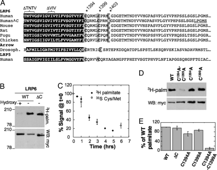
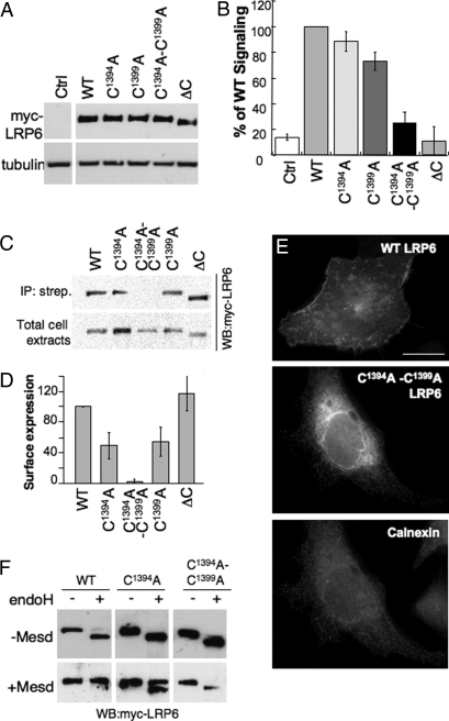
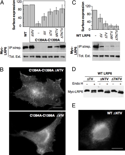
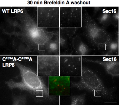
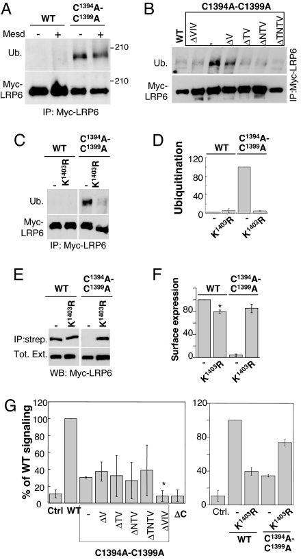
Canonical Wnt signaling is initiated by binding of Wnt proteins to members of the Frizzled family and subsequent complex formation with lipoprotein receptor-related proteins 5/6 (LRP5/6). Here, we show that LRP6 is palmitoylated on a juxtamembranous cysteine and that palmitoylation is required for exit from the endoplasmic reticulum (ER). We propose that palmitoylation serves to tilt the long, 23-residue transmembrane domain of LRP6 with respect to the plane of membrane to prevent a hydrophobic mismatch and subsequent recognition by the ER quality control. In support of this model, a palmitoylation-deficient LRP6 mutant could be rescued from ER retention by deletion of two to four residues in the transmembrane domain. Importantly, we found that palmitoylation-deficient LRP6 was retained in the ER by a completely novel monoubiquitination-dependent ER retention mechanism. Mutation of a specific lysine indeed abolished ubiquitination of palmitoylation-deficient LRP6 and led to a rescue from ER retention. Finally, at the cell surface, we found that interplay between palmitoylation and ubiquitination was necessary for efficient Wnt signaling.


Conflict of interest statement

The authors declare no conflict of interest.

Figures

Fig. 1.
Fig. 1.
LRP6 is palmitoylated during its entire lifetime. (A) Alignment of the TMD (black box) and the 47 first amino acids of the cytoplasmic tails of LRP6 and LRP5 from diverse species. LRP6ΔC corresponds to LRP6 with a truncated cytoplasmic tail that ends by the sequence LPGMS as underlined. ΔTNTV and ΔVIV indicate deletion mutants analyzed in Fig. 3. (B) HeLa cells transfected with myc-LRP6 or myc-LRP6ΔC were labeled with 3H-palmitic acid before immunoprecipitation. Immunoprecipitates were split into two, run on 4–12% SDS gels, and analyzed either by autoradiography or Western blotting (anti-myc). Chemical removal of S-palmitoylation was performed by treating cell extracts for 1 h at room temperature with 1M hydroxylamine hydrochloride, pH 7.2. (C) HeLa cells transfected with myc-LRP6 WT and Mesd were pulsed 30 min either with 3H-palmitic acid or 35S cysteine/methionine and chased for different times at 37°C in complete medium. Myc-LRP6 was immunoprecipitated and subjected to quantitative autoradiography. Results were normalized to the values at time 0. Error bars correspond to SD (n = 3). (D) HeLa cells transiently transfected with myc-LRP6 WT, C1394A, C1399A mutant or double mutant were incubated with 3H-palmitic acid before immunoprecipitation against Myc. Immunoprecipitates were split into two, run on 4–12% gels, and analyzed either by radiography or Western blotting (anti-myc). (E) Radioactive gels from D were quantified by using the Typhoon Imager. Values were first normalized with respect to the expression levels, and then mutants were normalized with respect to WT. Error bars correspond to SD (n = 4).
Fig. 2.
Fig. 2.
Palmitoylation of LRP6 is required for ER exit. (A and B) HEK293 cells were transfected with WT and mutants LRP6 and with Mesd. (A) The expression levels of WT and mutant LRP6 were determined by Western blotting against myc. (B) Wnt signaling was probed with the TOPFLASH reporter assay. The results are shown as mean normalized to WT. Error bars represent the SD (n = 4 experiments performed in triplicate). (C and D) HeLa cells transfected with plasmids encoding WT or mutant myc-tagged LRP6 and Mesd were submitted to surface biotinylation. (C) The levels of LRP6 in streptavidin immunoprecipitate (Upper) and in total cell lysate (Lower) were determined by quantitative Western blotting against myc. The amount of ΔV at the cell surface varied somewhat between experiments as indicated by the error bars and the absence of signal of the chosen blot. (D) Results were first normalized to the expression level and the mutants were normalized to WT. Error bars correspond to SD (n = 4). (E) HeLa cells expressing WT or mutant myc-LRP6 were analyzed by immunofluorescence against myc and calnexin. (Scale bar: 10 μM.) (F) HeLa cells were transfected 24 h with plasmids encoding WT or mutant myc-tagged LRP6 with and without Mesd. Forty micrograms of cell extracts was treated or not with endo H and analyzed by SDS/PAGE and Western blotting against myc.
Fig. 3.
Fig. 3.
Effect of TMD truncation on plasma membrane targeting of LRP6. (A) (Upper) HeLa cells were transfected 24 h with plasmids encoding Mesd and WT or palmitoylation-deficient myc-LRP6 mutants with deletions of one to four residues in their TMD (Fig. 1A): ΔTNTV, ΔNTV, ΔTV, ΔV, ΔVIV in the middle of the TMD. (Lower) The levels of LRP6 in the streptavidin immunoprecipitate and the total cell lysate were determined by quantitative anti-myc Western blotting. Error bars correspond to SD (n = 6 except for ΔTNTV, n = 4). Results were first normalized to the expression level and then to the surface expression of WT. (B) Localization of LRP6 mutants, coexpressed with Mesd, was analyzed by immunofluorescence. (Scale bar: 10 μm.) (C) (Upper) HeLa cells were transfected 24 h with plasmids encoding Mesd and LRP6 WT with deletions of zero to four residues in their TMD: ΔTNTV, ΔNTV, ΔTV, ΔV. (Lower) The levels of LRP6 in the streptavidin immunoprecipitate and the total cell lysate were determined by quantitative anti-myc Western blotting. Error bars correspond to SD (n = 3). (D) HeLa cells were transfected 24 h with the same plasmids as in C with and without Mesd. Forty micrograms of cell extracts were untreated or not with endo H and analyzed by SDS/PAGE and Western blotting against myc. (E) Localization of WT LRP6 ΔNTV, coexpressed with Mesd, was analyzed by immunofluorescence. (Scale bar: 10 μm.)
Fig. 4.
Fig. 4.
ER exit of WT and mutant LRP6. HeLa cells were transfected 24 h with plasmids encoding WT or mutant myc-LRP6 and cotransfected with Mesd. Cells were treated with brefeldin A for 1 h and then incubated in a brefeldin-free medium for 30 min at 37°C before fixation. Cells were analyzed by immunofluorescence against myc for LRP6 (green in the merged insets) and human sec16 (red in the merged insets). (Scale bar: 10 μM.)
Fig. 5.
Fig. 5.
ER retention of palmitoylation-deficient LRP6 is ubiquitin-mediated. (A–C) HeLa cells were transfected 24 h with plasmids encoding WT or mutant myc-LRP6 and cotransfected or not with Mesd. Immunoprecipitates against myc-LRP6 were analyzed by SDS/PAGE and Western blotting against ubiquitin and myc. (D) The levels of WT and mutant LRP6 in the streptavidin immunoprecipitate and the total cell lysat from C were quantified with a Typhoon imager and normalized to that of the C1394A-C1399A-LRP6 mutant. Error bars correspond to SD (n = 3). (E and F) HeLa cells were transfected 24 h with plasmids encoding Mesd and WT or myc-LRP6 mutants. The levels of LRP6 in the streptavidin immunoprecipitate and the total cell lysate were determined by quantitative anti-myc Western blotting. Error bars correspond to SD (n = 4). Results were first normalized to the expression level and then to the surface expression of WT. (G) WT or mutant myc-tagged LRP6 cotransfected with Mesd into HEK293 cells were tested by using the TOPFLASH reporter assay. The results are shown as mean normalized to WT and standard deviations of the mean of three independent experiments done in triplicate. In Left none of the TMD truncation mutants were significantly different from the full-length C1394A-C1399A mutant except the ΔVIV mutant, which was significantly more deficient in signaling (*, P < 0.005).

Similar articles

Cited by

References

    1. Cadigan KM, Nusse R. Wnt signaling: A common theme in animal development. Genes Dev. 1997;11:3286–3305. - PubMed
    1. Kikuchi A, Yamamoto H, Kishida S. Multiplicity of the interactions of Wnt proteins and their receptors. Cell Signal. 2007;19:659–671. - PubMed
    1. Clevers H. Wnt/β-catenin signaling in development and disease. Cell. 2006;127:469–480. - PubMed
    1. Koduri V, Blacklow SC. Requirement for natively unstructured regions of mesoderm development candidate 2 in promoting low-density lipoprotein receptor-related protein 6 maturation. Biochemistry. 2007;46:6570–6577. - PubMed
    1. Hsieh JC, et al. Mesd encodes an LRP5/6 chaperone essential for specification of mouse embryonic polarity. Cell. 2003;112:355–367. - PubMed

Publication types

Substances

LinkOut - more resources