. 2008 Apr 8;105(14):5519-24.
doi: 10.1073/pnas.0801779105. Epub 2008 Mar 31.

Tbx5-dependent pathway regulating diastolic function in congenital heart disease

Affiliations

Tbx5-dependent pathway regulating diastolic function in congenital heart disease

Yonghong Zhu et al. Proc Natl Acad Sci U S A. .

Abstract

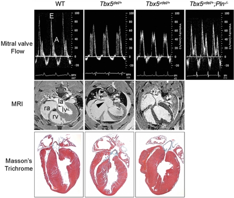
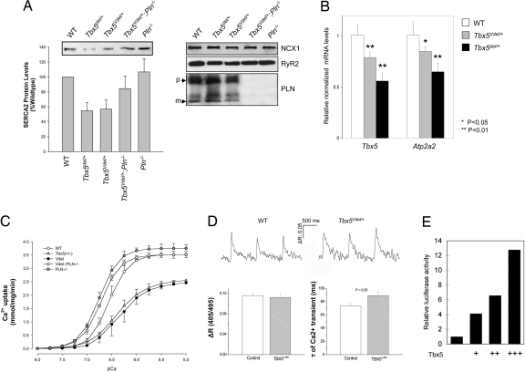
At the end of every heartbeat, cardiac myocytes must relax to allow filling of the heart. Impaired relaxation is a significant factor in heart failure, but all pathways regulating the cardiac relaxation apparatus are not known. Haploinsufficiency of the T-box transcription factor Tbx5 in mouse and man causes congenital heart defects (CHDs) as part of Holt-Oram syndrome (HOS). Here, we show that haploinsufficiency of Tbx5 in mouse results in cell-autonomous defects in ventricular relaxation. Tbx5 dosage modulates expression of the sarco(endo)plasmic reticulum Ca(2+)-ATPase isoform 2a encoded by Atp2a2 and Tbx5 haploinsufficiency in ventricular myocytes results in impaired Ca(2+) uptake dynamics and Ca(2+) transient prolongation. We also demonstrate that Tbx5 can activate the Atp2a2 promoter. Furthermore, we find that patients with HOS have significant diastolic filling abnormalities. These results reveal a direct genetic pathway that regulates cardiac diastolic function, implying that patients with structural CHDs may have clinically important underlying anomalies in heart function that merit treatment.


Conflict of interest statement

The authors declare no conflict of interest.

Figures

Fig. 1.
Fig. 1.
Diastolic dysfunction in Tbx5 ventricle-specific haploinsufficient mice. (Top) Doppler signal at the MV showing E and A waves. The E wave is decreased, and the A wave is increased in Tbx5del/+ and in Tbx5Vdel/+ mice. These values are restored to normal in Tbx5Vdel/+;PLN−/− mice. (Middle) MRI showing ASDs (arrowhead) in Tbx5del/+ but not in Tbx5Vdel/+ mice. la, left atrium; lv, left ventricle; ra, right atrium; rv, right ventricle. (Bottom) Histology of adult hearts stained by Masson's trichrome.
Fig. 2.
Fig. 2.
Tbx5 modulates SERCA2 expression. (A) Western blots showing decreased levels of SERCA2 protein in Tbx5del/+ and Tbx5Vdel/+ ventricular extracts. Normal levels of Ncx1, RyR2, and PLN were found in all genotypes. (B) Quantitative RT-PCR demonstrating decreased Tbx5 and Atp2a2 mRNA levels in Tbx5del/+ and Tbx5Vdel/+ ventricular extracts (n = 6 for each genotype; means ± SEMs are shown). *, P < 0.05; **, P < 0.01. (C) Ca2+ uptake in ventricular tissues demonstrating impaired Ca2+ uptake in Tbx5del/+ and Tbx5Vdel/+ mice and restoration to WT levels in Tbx5Vdel/+;PLN−/− mice. (D) Intracellular Ca2+ transients. (Upper) Representative Ca2+ transient recordings obtained from control and Tbx5Vdel/+ cardiomyocytes under field stimulation at 1 Hz (35.5 ± 0.5°C) and loaded with indo 1-AM. ΔR stands forΔR405/495, the ratio of the intensity of fluorescence emitted at 405 nm to that at 495 nm, which is used as an index of intracellular Ca2+ concentrations in this study. (Lower) Mean data of Ca2+-transient amplitude (ΔR405/495) and time constant (τ) of Ca2+ transient decay (in milliseconds) obtained from myocytes isolated from control (n = 41 cells) and Tbx5Vdel/+ (n = 43 cells) mice. (E) Luciferase reporter assay shows dosage-dependent activation of Atp2a2-luciferase. Expression vectors: +, 100 ng; ++, 250 ng; +++, 500 ng.
Fig. 3.
Fig. 3.
Doppler ultrasound shows diastolic dysfunction in patients with HOS. Measurements are made from the four-chamber view; the pulsed Doppler sample is placed on the MV orifice, the MV annulus, and the IVS. The MV inflow pattern (Top) and tissue Doppler patterns for the MV (Middle) and IVS (Bottom) are shown in a patient with HOS (Right) compared with an age-matched control (Left). All measurements are in ms. The MV inflow pattern shows a larger E wave in the patient with HOS; tissue velocities for the MV and IVS tissue also are lower. Compared with the age-matched control, this indicates a more restrictive pattern of filling.

Similar articles

Cited by

References

    1. Kass DA, Bronzwaer JG, Paulus WJ. What mechanisms underlie diastolic dysfunction in heart failure? Circ Res. 2004;94:1533–1542. - PubMed
    1. MacLennan DH, Kranias EG. Phospholamban: A crucial regulator of cardiac contractility. Nat Rev Mol Cell Biol. 2003;4:566–577. - PubMed
    1. Maclennan DH. Interactions of the calcium ATPase with phospholamban and sarcolipin: Structure, physiology and pathophysiology. J Muscle Res Cell Motil. 2004;25:600–601. - PubMed
    1. Dillmann WH. Regulation of expression of cardiac sarcoplasmic reticulum proteins under pathophysiological conditions. Mol Cell Biochem. 1996;157:125–128. - PubMed
    1. Srivastava D. Making or breaking the heart: From lineage determination to morphogenesis. Cell. 2006;126:1037–1048. - PubMed

Publication types

MeSH terms

Substances

Grants and funding