Comparative Study
. 2008 Apr;67(4):309-18.
doi: 10.1097/NEN.0b013e31816a1df3.

Cholinergic neuronal and axonal abnormalities are present early in aging and in Alzheimer disease

Affiliations
Comparative Study

Cholinergic neuronal and axonal abnormalities are present early in aging and in Alzheimer disease

Changiz Geula et al. J Neuropathol Exp Neurol. 2008 Apr.

Abstract

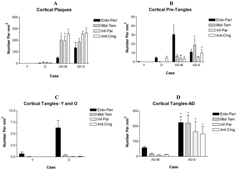
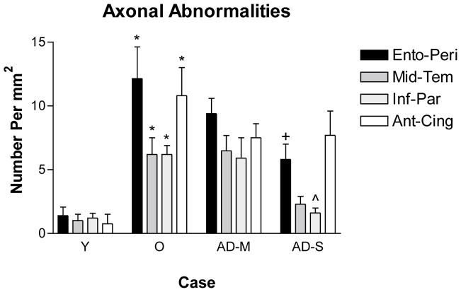
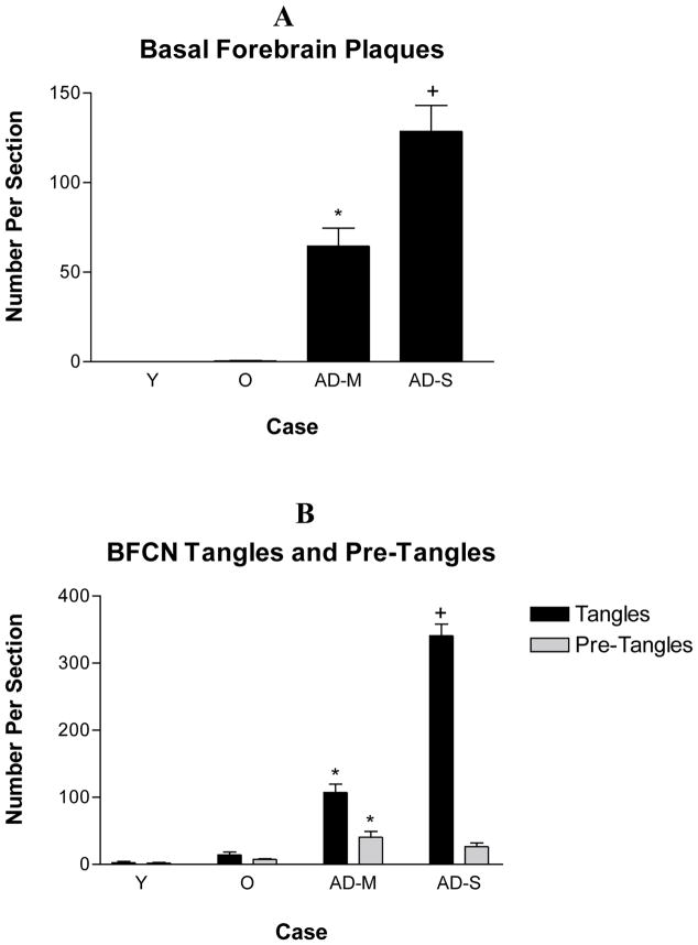
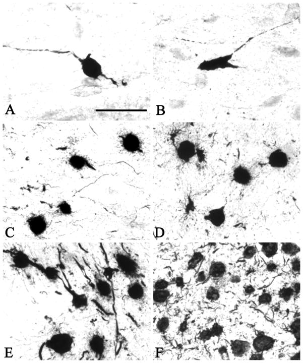
A large body of evidence indicates that basal forebrain cholinergic neurons are selectively vulnerable to degeneration early in Alzheimer disease (AD). Recent studies, however, demonstrate reductions in cortical activity of the cholinergic enzyme choline acetyltransferase only in late stages of AD. To address this apparent contradiction, we compared abnormalities in magnocellular basal forebrain cholinergic neurons and their axons in nondemented young (<65 years; n = 6), nondemented old (>65 years; n = 7), pathologically mild (n = 5), and pathologically severe (n = 5) AD cases. Cholinergic axon abnormalities (i.e. thickened fibers and ballooned terminals) were evident in nondemented middle-aged cases, increased in nondemented old cases, and reduced in density in severe AD. This suggests that loss of cortical cholinergic axons in AD occurs preferentially in fibers with these abnormalities. Paired helical filament 1-immunoreactive pretangles and tangles were observed as early as the third decade prior to their appearance in entorhinal/perirhinal cortex; they were increased in mild and severe AD. These results indicate that basal forebrain cholinergic neuron abnormalities are present very early in aging and in the course of AD. Therefore, despite the morphologic alterations, choline acetyltransferase activity, but not necessarily normal neuron functions, may be preserved.


Figures

Figure 1
Figure 1
The density of cortical plaques and tangles in the four groups of subjects. The only pathology observed in young cases (<65 years) consisted of a small number of pre-tangles and tangles in the entorhinal/peri-rhinal cortex. The density of plaques, tangles and pre-tangles displayed a progressive decrease in non-demented old and AD cases. Based on the relative density of cortical plaques and tangles and the Braak staging of tangle formation (Table 1), AD cases were divided into pathologically mild and pathologically severe cases. The density of tangles in all cortical areas and the density of plaques in the entorhinal cortex were statistically significantly greater in pathologically severe AD when compared with pathologically mild AD. A. * p<0.001 compared to normal old, +p<0.01 compared with pathologically mild. B. * and + p<0.05 compared with non-demented old. D. * p<0.001 compared with non-demented old, p<0.01 compared with pathologically mild. Analysis was carried out using all cases in Table 1 except those identified by an asterisk. Y – Non-Demented Young, O – Non-Demented Old, AD-M, Pathologically Mild AD, AD-S – Pathologically Severe AD.
Figure 2
Figure 2
In the youngest case investigated (Case #1), AChE activity (A) and ChAT immunoreactivity (B) were present within cholinergic axons homogenously thin in diameter. Immunohistochemistry for ChAT visualized small varicosities within axons. In brains from older cases and AD brains, one or more of a number of abnormalities were seen in both AChE (C, E and G) and ChAT (D, F, H) stained material. These included thickened axons (C and D) and swollen and ballooned varicosities at axonal endings (E–H), which often occurred in a chandelier arrangement. Scale bar in A is 100 μm and also applies to B, and the scale bar in C is 50 μm and also applies to D–H.
Figure 3
Figure 3
The density of cortical cholinergic axonal abnormalities in the four groups of subjects. No such abnormalities were present in the youngest cases (Case # 1 and 2). A small number of such abnormalities were present in the rest of the cases below 64 years and the density of such abnormalities displayed a significant increase in non-demented old cases compared with the young (p<0.05). The density of axonal abnormalities remained constant in pathologically mild AD and was reduced in pathologically severe AD and significantly so in the entorhinal cortex and the inferior parietal lobule. Quantitative analysis was conducted on all of the cases in Table 1 except those identified by an asterisk. Y – Non-Demented Young, O – Non-Demented Old, AD-M, Pathologically Mild AD, AD-S – Pathologically Severe AD.
Figure 4
Figure 4
The number of basal forebrain plaques, tangles and pre-tangles within cholinergic neurons in each section. Plaques were absent from the basal forebrain of young individuals. They were seen in old individuals in moderate numbers. A significant increase in the number of plaques was seen in pathologically mild AD and another significant increase in pathologically severe AD. The density of pre-tangles was higher in pathologically mild AD when compared with non-demented old cases. The density of tangles showed a further significant increase in the cholinergic neurons in pathologically severe AD. Quantitative analysis was carried out using all of the cases in Table 1 except those identified by an asterisk. A. *p<0.001 compared with non-demented old cases, +p<0.001 compared with pathologically mild cases. B. *p<0.001 compared with non-demented old cases, +p<0.001 compared with pathologically mild cases. Y – Non-Demented Young, O – Non-Demented Old, AD-M, Pathologically Mild AD, AD-S – Pathologically Severe AD.
Figure 5
Figure 5
PHF1-positive pre-tangles and tangles in the basal forebrain of young (A and B, Case #1), middle aged (C, Case #), old (D, Case #), pathologically mild (E, Case #) and pathologically severe AD (F, Case #). Pre-tangles were present in the cholinergic neurons in the 26 year-old (Case 1, A and B) in whose brain no tangles and pre-tangles were found in the entorhinal/peri-rhinal cortex. Thereafter, the numbers of tangles and pre-tangles within the basal forebrain cholinergic neurons displayed a progressive increase. Scale bar in A is 100 μm and also applies to B-F.

References

    1. Arendash GW, Millard WJ, Dunn AJ, Meyer EM. Long-term neuropathological and neurochemical effects of nucleus basalis lesions in the rat. Science. 1987;238:952–6. - PubMed
    1. Arendt T, Bigl V, Tennstedt A, Arndt A. Neuronal loss in different parts of the nucleus basalis is related to neuritic plaque formation in cortical target areas in Alzheimer’s disease. Neurosci. 1985;14:1–14. - PubMed
    1. Arnold SE, Hyman BT, Flory J, Damasio AR, Van Hoesen GW. The topographical and neuroanatomical distribution of neurofibrillary tangles and neuritic plaques in the cerebral cortex of patients with Alzheimer’s disease. Cerebral Cortex. 1991;1:103–16. - PubMed
    1. Arriagada PV, Marzloff K, Hyman BT. Distribution of Alzheimer-type pathologic changes in nondemented elderly individuals matches the pattern in Alzheimer’s disease. Neurology. 1992;42:1681–8. - PubMed
    1. Bierer LM, Haroutunian V, Gabriel S, Knott PJ, Carlin LS, Purohit DP, Schmeidler J, Kanof P, Davis KL. Neurochemical correlates of dementia severity in Alzheimer’s disease: relative importance of the cholinergic deficits. J Neurochem. 1995;64:749–50. - PubMed

Publication types

MeSH terms