

Perfusion MR imaging for differentiation of benign and malignant meningiomas
- PMID: 18379768
- PMCID: PMC2440923
- DOI: 10.1007/s00234-008-0373-y
Perfusion MR imaging for differentiation of benign and malignant meningiomas
Abstract
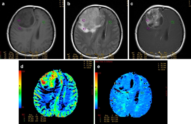
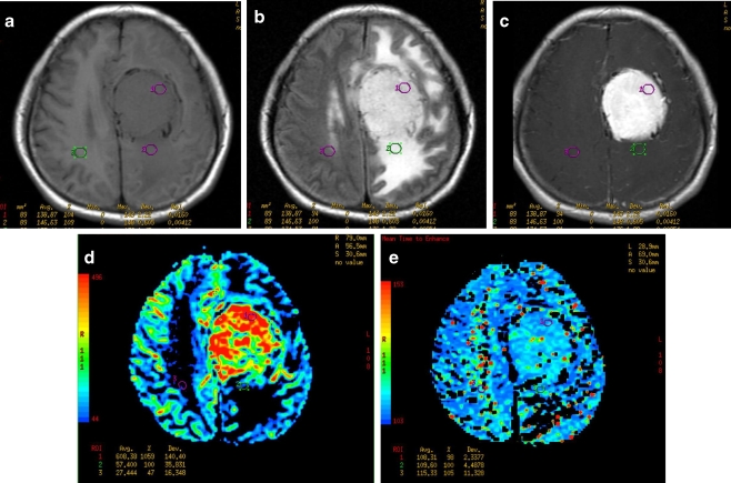
Introduction: Our purpose was to determine whether perfusion MR imaging can be used to differentiate benign and malignant meningiomas on the basis of the differences in perfusion of tumor parenchyma and/or peritumoral edema.
Methods: A total of 33 patients with preoperative meningiomas (25 benign and 8 malignant) underwent conventional and dynamic susceptibility contrast perfusion MR imaging. Maximal relative cerebral blood volume (rCBV) and the corresponding relative mean time to enhance (rMTE) (relative to the contralateral normal white matter) in both tumor parenchyma and peritumoral edema were measured. The independent samples t-test was used to determine whether there was a statistically significant difference in the mean rCBV and rMTE ratios between benign and malignant meningiomas.
Results: The mean maximal rCBV values of benign and malignant meningiomas were 7.16+/-4.08 (mean+/-SD) and 5.89+/-3.86, respectively, in the parenchyma, and 1.05+/-0.96 and 3.82+/-1.39, respectively, in the peritumoral edema. The mean rMTE values were 1.16+/-0.24 and 1.30+/-0.32, respectively, in the parenchyma, and 0.91+/-0.25 and 1.24+/-0.35, respectively, in the peritumoral edema. The differences in rCBV and rMTE values between benign and malignant meningiomas were not statistically significant (P>0.05) in the parenchyma, but both were statistically significant (P<0.05) in the peritumoral edema.
Conclusion: Perfusion MR imaging can provide useful information on meningioma vascularity which is not available from conventional MRI. Measurement of maximal rCBV and corresponding rMTE values in the peritumoral edema is useful in the preoperative differentiation between benign and malignant meningiomas.
Figures
References
-
- Kleihues P, Louis DN, Scheithauer BW, Rorke LB, Reifenberger G, Burger PC, Cavenee WK. The WHO classification of tumors of the nervous system. J Neuropathol Exp Neurol. 2002;61:215–225. - PubMed
-
- Brainard JA, Prayson RA, Barnett GH. Frozen section evaluation of stereotactic brain biopsies: diagnostic yield at the stereotactic target position in 188 cases. Arch Pathol Lab Med. 1997;121:481–484. - PubMed
-
- Knopp EA, Cha S, Johnson G, Mazumdar A, Golfonos JG, Zagzag D, Kelly PJ, Kricheff II. Glial neoplasms: dynamic contrast-enhanced T2*-weighted MR imaging. Radiology. 1999;211:791–798. - PubMed
Publication types
MeSH terms
Substances
LinkOut - more resources
Full Text Sources
