

Mechanism of light-induced translocation of arrestin and transducin in photoreceptors: interaction-restricted diffusion
- PMID: 18379987
- PMCID: PMC2717607
- DOI: 10.1002/iub.7
Mechanism of light-induced translocation of arrestin and transducin in photoreceptors: interaction-restricted diffusion
Abstract
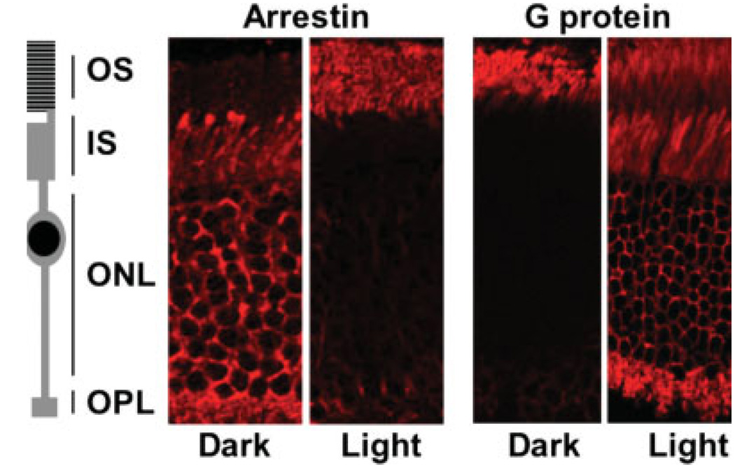
Many signaling proteins change their location within cells in response to external stimuli. In photoreceptors, this phenomenon is remarkably robust. The G protein of rod photoreceptors and rod transducin concentrates in the outer segments (OS) of these neurons in darkness. Within approximately 30 minutes after illumination, rod transducin redistributes throughout all of the outer and inner compartments of the cell. Visual arrestin concurrently relocalises from the inner compartments to become sequestered primarily within the OS. In the past several years, the question of whether these proteins are actively moved by molecular motors or whether they are redistributed by simple diffusion has been extensively debated. This review focuses on the most essential works in the area and concludes that the basic principle driving this protein movement is diffusion. The directionality and light dependence of this movement is achieved by the interactions of arrestin and transducin with their spatially restricted binding partners.
Figures
References
-
- Arshavsky VY, Lamb TD, Pugh EN., Jr G proteins and phototransduction. Annu. Rev. Physiol. 2002;64:153–187. - PubMed
-
- Fain GL, Matthews HR, Cornwall MC, Koutalos Y. Adaptation in vertebrate photoreceptors. Physiol. Rev. 2001;81:117–151. - PubMed
-
- Makino CL, Wen XH, Lem J. Piecing together the timetable for visual transduction with transgenic animals. Curr. Opin. Neurobiol. 2003;13:404–412. - PubMed
-
- Broekhuyse RM, Tolhuizen EF, Janssen AP, Winkens HJ. Light induced shift and binding of S-antigen in retinal rods. Curr. Eye Res. 1985;4:613–618. - PubMed
-
- Philp NJ, Chang W, Long K. Light-stimulated protein movement in rod photoreceptor or cells of the rat retina. FEBS Lett. 1987;225:127–132. - PubMed
Publication types
MeSH terms
Substances
Grants and funding
LinkOut - more resources
Full Text Sources
Miscellaneous
