Review
. 2008 Nov;1779(11):682-91.
doi: 10.1016/j.bbagrm.2008.03.001. Epub 2008 Mar 12.

MicroRNA-206: the skeletal muscle-specific myomiR

Affiliations
Review

MicroRNA-206: the skeletal muscle-specific myomiR

John J McCarthy. Biochim Biophys Acta. 2008 Nov.

Abstract

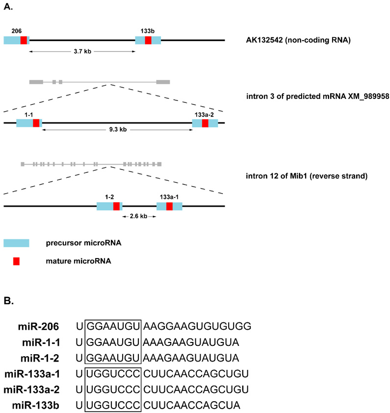
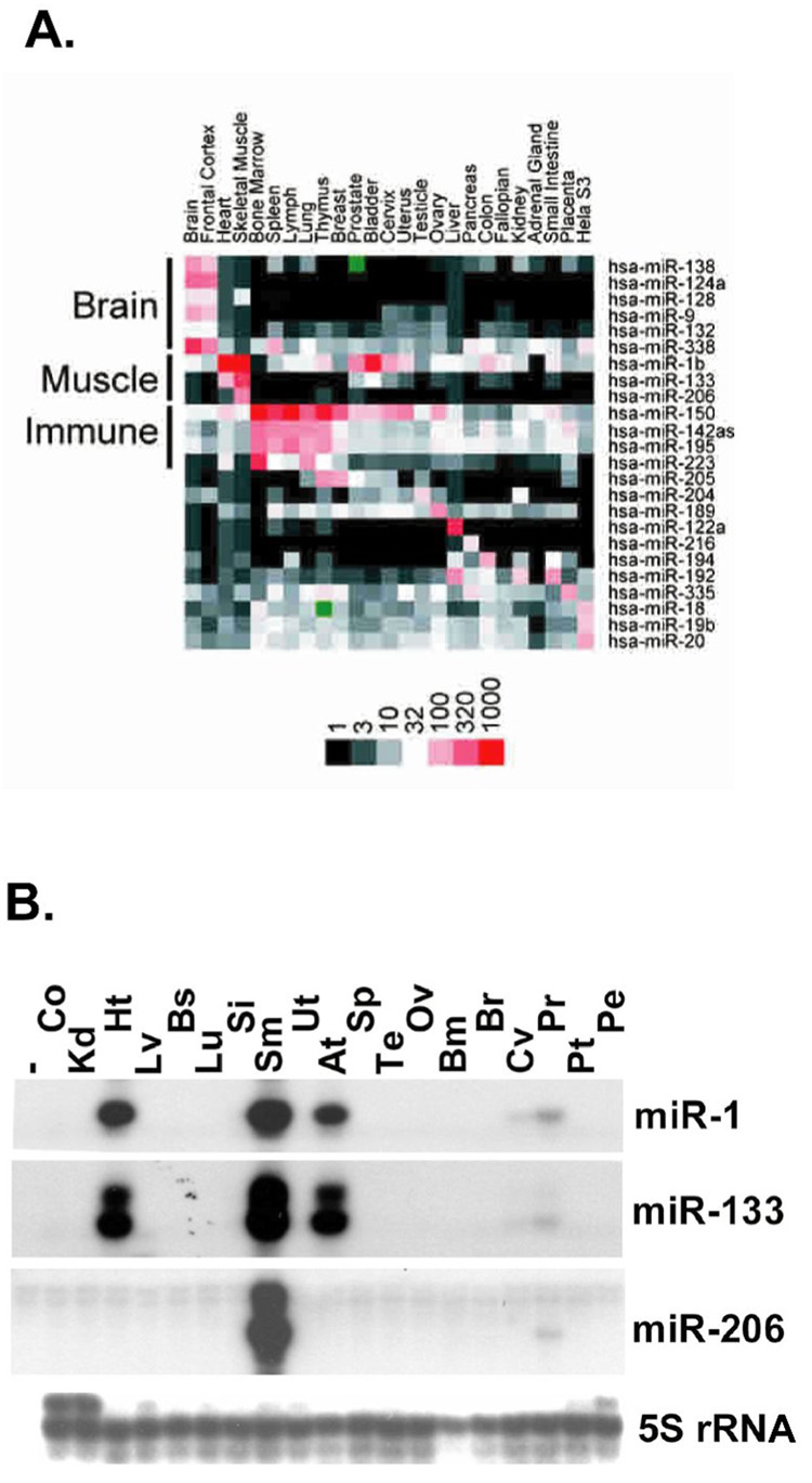
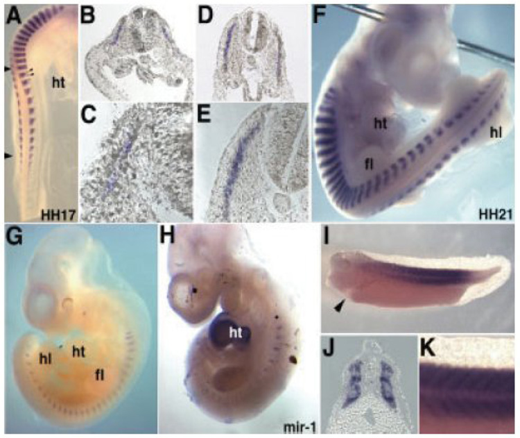
MicroRNAs (miRNAs) are a class of non-coding RNAs involved in post-transcriptional gene silencing. A small number of striated muscle-specific miRNAs have been identified and shown to have an important role in myogenesis, embryonic muscle growth and cardiac function and hypertrophy. One of these myomiRs (myo=muscle+miR=miRNA), miR-206, is unique in that it is only expressed in skeletal muscle. The purpose of this review is to discuss what is currently known about miR-206 and its function in myogenesis as well as propose potential new roles for miR-206 in skeletal muscle biology. The review is also intended to serve as a comprehensive resource for miR-206 with the hope of encouraging further research on the role of miR-206 in skeletal muscle.


Figures

Figure 1
Figure 1. Muscle-specific miRNAs
A, the location of mouse precursor and mature miRNA within the primary miRNA transcript for each of the muscle-specific gene clusters: miR-206 and miR-133b are processed from the non-coding RNA AK132542A; miR-1-1 and miR-133a-2 are derived from intron three of the predicted mRNA XM_989958; miR-1-2 and miR-133a-1 are encoded in the 12th intron of the mindbomb 1 transcript on the reverse strand. B, Sequence alignment of each muscle-specific miR. The “seed” region is boxed for the miR-1/206 and the miR-133a/b groups to emphasize their respective conservation. Each gene cluster consists of a member from each of these subgroups.
Figure 2
Figure 2. Skeletal muscle-specific expression of miR-206
A, MicroRNA expression profiling of various human tissues identify the myomiRs (miR-1, -133a and -206) and show miR-206 expression is restricted to skeletal muscle. Image is reproduced from Baskerville and Bartel (2005) with permission granted from Cold Spring Harbor Laboratory Press [14]. B. RNase protection assay of different human tissues clearly shows that miR-206 is skeletal muscle specific, confirming microarray analysis presented in A; Co, colon; Kd, kidney; Ht, heart; Lv, liver; Bs, breast; Lu, lung; Si, small intestine; Sm, skeletal muscle; Ut, uterus; At, atrium; Sp, spleen; Te, testis; Ov, ovary; Bm, bone marrow; Br, brain; Cv, cervix; Pt, prostate tumor; Pe, prostate epithelium. The image is reproduced from Kim et al (2006) with permission from Rockefeller University Press [37].
Figure 3
Figure 3. Expression of miR-206 is restricted to somites in mouse, chick and frog embryos
A, miR-206 expression in somite of chick embryo (stage HH17) with small arrows at axial level indicating expression in lateral myotome; B–E, different magnifications of transverse sections through trunk of chick embryo (stage HH17) reveal miR-206 expression in myotome; F, mir-206 expression in chick embryo at stage HH21; G, somite-specific expression of miR-206 mouse embryo at E10; H, in contrast to miR-206, miR-1 is expressed in the heart and somites in the mouse embryo at E10; I-K, expression of miR-206 in Xenopus laevis tadpole (stage 35) is restricted to somites with arrow in (K) indicating lack of expression in heart, (J) transverse section through trunk show somite staining and (K) is higher magnification of (I); fl; forelimb, ht, heart; hl, hind limb. Image is reproduced from Sweetman et al (2006) with permission from John Wiley & Sons, Inc. [48].
Figure 4
Figure 4. Promoter of miRNA-206 gene
A, a screen capture from CisView schematically displays characterization of upstream sequences of miRNA-206 gene. Within this presumptive promoter sequence (~ 1kb) two highly conserved cis-regulatory modules (CRM) are identified that are separated by a stretch of low complexity repeats. B, nucleotide sequence of miRNA-206 promoter with the two regulatory modules, CRM-1 and CRM-2, as identified by CisView boxed. Highlighted (red) is the TATA-box in CRM-1 and the three Myod1 E-boxes located in CRM-2. Using an in vitro model, preliminary data revealed CRM-2 is sufficient for miRNA-206 up-regulation during myogenic differentiation.
Figure 5
Figure 5. MiRNA regulation of circadian rhythms in skeletal muscle
A diagram of a proposed model in which miR-206 is involved in a negative feedback loop that regulates Clock expression. Evidence to support the model are as follows; Expression profiling identified Myod1 as a clock-controlled gene with a circadian pattern of expression [114]. In turn, Myod1 has been shown to drive expression of primary miRNA-206 transcript AK132542 leading to increased expression of miR-206 [52]. MiRNA-206 is predicted to target Clock gene by three different databases (TargetScan, Miranda and PicTar). The model provides a novel mechanism by which skeletal muscle-specific factors (Myod1 and miR-206) can regulate circadian rhythms in a tissue-specific manner.

References

    1. Pasquinelli AE, Reinhart BJ, Slack F, Martindale MQ, Kuroda MI, Maller B, Hayward DC, Ball EE, Degnan B, Muller P, Spring J, Srinivasan A, Fishman M, Finnerty J, Corbo J, Levine M, Leahy P, Davidson E, Ruvkun G. Conservation of the sequence and temporal expression of let-7 heterochronic regulatory RNA. Nature. 2000;408:86–89. - PubMed
    1. Arasu P, Wightman B, Ruvkun G. Temporal regulation of lin-14 by the antagonistic action of two other heterochronic genes, lin-4 and lin-28. Genes Dev. 1991;5:1825–1833. - PubMed
    1. Wightman B, Burglin TR, Gatto J, Arasu P, Ruvkun G. Negative regulatory sequences in the lin-14 3'-untranslated region are necessary to generate a temporal switch during Caenorhabditis elegans development. Genes Dev. 1991;5:1813–1824. - PubMed
    1. Wightman B, Ha I, Ruvkun G. Posttranscriptional regulation of the heterochronic gene lin-14 by lin-4 mediates temporal pattern formation in C. elegans. Cell. 1993;75:855–862. - PubMed
    1. Ha I, Wightman B, Ruvkun G. A bulged lin-4/lin-14 RNA duplex is sufficient for Caenorhabditis elegans lin-14 temporal gradient formation. Genes Dev. 1996;10:3041–3050. - PubMed

Publication types

LinkOut - more resources