Review
. 2008 Jul 6;5(24):671-90.
doi: 10.1098/rsif.2008.0052.

Optical tweezers for single cells

Affiliations
Review

Optical tweezers for single cells

Hu Zhang et al. J R Soc Interface. .

Abstract

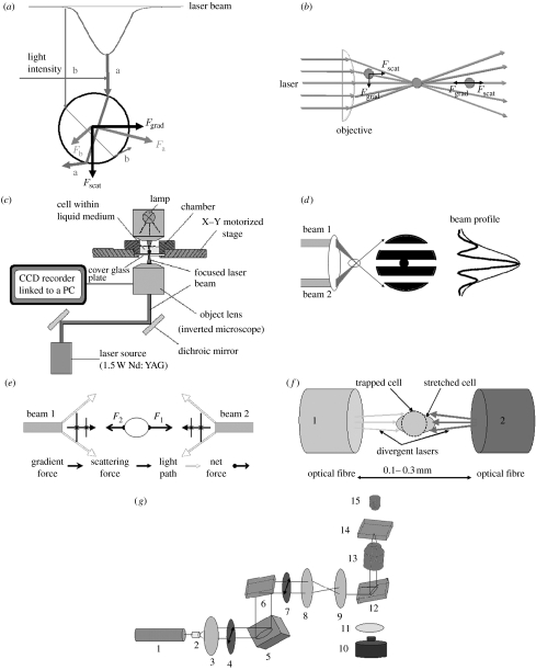
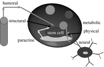
Optical tweezers (OT) have emerged as an essential tool for manipulating single biological cells and performing sophisticated biophysical/biomechanical characterizations. Distinct advantages of using tweezers for these characterizations include non-contact force for cell manipulation, force resolution as accurate as 100aN and amiability to liquid medium environments. Their wide range of applications, such as transporting foreign materials into single cells, delivering cells to specific locations and sorting cells in microfluidic systems, are reviewed in this article. Recent developments of OT for nanomechanical characterization of various biological cells are discussed in terms of both their theoretical and experimental advancements. The future trends of employing OT in single cells, especially in stem cell delivery, tissue engineering and regenerative medicine, are prospected. More importantly, current limitations and future challenges of OT for these new paradigms are also highlighted in this review.


Figures

Figure 1
Figure 1
Working principles of optical tweezers. (a) The scattering (Fscat) and gradient (Fgrad) components of optical forces on a dielectric sphere due to a Gaussian laser beam (light intensity increases from b to a). (b) Schematic of optical tweezers. Light enters the objective lens of a microscope and is focused to a diffraction-limited beam waist, creating a three-dimensional light gradient; a particle that is out of trap is brought back to the centre of the trap. (c) Typical experimental set-up of optical tweezers, adapted from the technical note of Cell Robotic Inc., USA. (d) Two-beam interferometric optical tweezers, adapted from Chiou et al. (1997). (e) Schematic of gradient and scattering forces for two beams, adapted from Constable et al. (1993). (f) Schematic of optical stretcher, adapted from Guck et al. (2000). (g) Schematic of holographic optical tweezers generated from a spatial light modulator (SLM), adapted from Martin-Badosa et al. (2007). 1, laser beam; 2, LC lens; 3, LE lens; 4, polarizing element; 5, SLM; 6, dichroic mirror; 7, polarizing element; 8, telescope; 9, telescope; 10, CCD camera; 11, tube lens; 12, dichroic mirror; 13, microscopic objective; 14, sample; 15, illumination.
Figure 2
Figure 2
Optical tweezers for cell sorting. Targeted cells are detected in the detection region and identification of those cells triggers switching on either multiple traps or single trap. Cells are driven by optical tweezers to be delivered to the collection output while all other cells flow to the waste output. Adapted from Wang et al. (2005).
Figure 3
Figure 3
Stretching RBCs by optical tweezers. (a) Two diametrically opposed silica beads of 4.1 μm are attached onto an RBC surface. (b) One bead is trapped by optical tweezers while the other is fixed onto a glass surface. Deformation is achieved by moving the glass surface to the opposite direction. (c) Large deformations of RBCs in phosphate buffer saline solution at room temperature are captured by optical micrographs under different trapping forces. Adapted from Lim et al. (2004a).
Figure 4
Figure 4
Stem cell niche. Elements are identified for regulating the system of a stem cell, including the constraints of the architectural space, physical engagement of the cell membrane, signalling interactions, neural input and metabolic products of tissue activity. Adapted from Scadden (2006).

References

    1. Abbondanzieri E.A, Greenleaf W.J, Shaevitz J.W, Landick R, Block S.M. Direct observation of base-pair stepping by RNA polymerase. Nature. 2005;438:460–465. doi: 10.1038/nature04268. - DOI - PMC - PubMed
    1. Akselrod G.M, Timp W, Mirsaidov U, Zhao Q, Li C, Timp K, Matsudaira P, Timp G. Laser-guided assembly of heterotypic three-dimensional living cell microarrays. Biophys. J. 2006;91:3465–3473. doi: 10.1529/biophysj.106.084079. - DOI - PMC - PubMed
    1. Allemand J.F, Bensimon D, Croquette V. Stretching DNA and RNA to probe their interactions with proteins. Curr. Opin. Struct. Biol. 2003;13:266–274. doi: 10.1016/S0959-440X(03)00067-8. - DOI - PubMed
    1. Allen L, Beijersbergen M.W, Spreeuw R.J.C, Woerdman J.P. Orbital angular momentum of light and the transformation of laguerre-gaussian laser modes. Phys. Rev. A. 1992;45:8185–8189. doi: 10.1103/PhysRevA.45.8185. - DOI - PubMed
    1. Andersson M, Madgavkar A, Stjerndahl M, Wu Y, Tan W, Duran R, Niehren S, Mustafa M, Arvidson K. Using optical tweezers for measuring the interaction forces between human bone cells and implant surfaces: system design and force calibration. Rev. Sci. Instrum. 2007a;78:074302. doi: 10.1063/1.2752606. - DOI - PubMed

Publication types

MeSH terms

LinkOut - more resources