. 2008 Jun 13;283(24):16950-9.
doi: 10.1074/jbc.M800783200. Epub 2008 Mar 31.

Functional surfaces on the p35/ARPC2 subunit of Arp2/3 complex required for cell growth, actin nucleation, and endocytosis

Affiliations

Functional surfaces on the p35/ARPC2 subunit of Arp2/3 complex required for cell growth, actin nucleation, and endocytosis

Karen M Daugherty et al. J Biol Chem. .

Abstract

The Arp2/3 complex is comprised of seven evolutionarily conserved subunits and upon activation by WASp or another nucleation promoting factor nucleates the formation of actin filaments. These events are critical for driving a wide range of cellular processes, including motility, endocytosis, and intracellular trafficking. However, an in depth understanding of the Arp2/3 complex activation and nucleation mechanism is still lacking. Here, we used a mutagenesis approach in Saccharomyces cerevisiae to dissect the structural and functional roles of the p35/ARPC2 subunit. Using integrated alleles that target conserved and solvent-exposed residues, we identified surfaces on p35/ARPC2 required for cell growth, actin organization, and endocytosis. In parallel, we purified the mutant Arp2/3 complexes and compared their actin assembly activities both in the presence and in the absence of WASp. The majority of alleles with defects mapped to one face of p35/ARPC2, where there was a close correlation between loss of actin nucleation and endocytosis. A second site required for nucleation and endocytosis was identified near the contact surface between p35/ARPC2 and p19/ARPC4. A third site was identified at a more distal conserved surface, which was critical for endocytosis but not nucleation. These findings pinpoint the key surfaces on p35/ARPC2 required for Arp2/3 complex-mediated actin assembly and cellular function and provide a higher resolution view of Arp2/3 structure and mechanism.


Figures

FIGURE 1.
FIGURE 1.
Scanning mutagenesis of conserved surface residues on the p35/ARPC2 subunit of Arp2/3 complex. A, alignment of p35/ARPC2 sequences from S. cerevisiae (S.c.) and B. taurus (B.t.). Mutant arc35 allele numbers are color-coded according to cell growth phenotypes: pseudo wild type, green; mild ts, yellow; strong ts, orange; lethal, red. Black dots denote conserved residues that are surface exposed on the crystal structure of bovine Arp2/3 complex (3). B, positions of residues mutated in each arc35 allele mapped on the bovine Arp2/3 complex structure (3). The p35/ARPC2 subunit is tan, and each subunit is labeled.
FIGURE 2.
FIGURE 2.
Cell growth phenotypes of arc35 mutants. A, strains carrying the indicated arc35 alleles on low copy (CEN) LEU2 plasmids were transformed into an arc35Δ strain covered by a URA3-marked ARC35 plasmid. Transformants were selected on Leu- medium and then plated on 5-FOA to counter-select against URA3. B, dissection of lethal arc35-105 and arc35-106 alleles. Residues mutated (alanine substitutions) in each new allele are indicated, and growth phenotypes of each allele (Fig. 2A and 2C) are listed. C, nonlethal integrated arc35 mutant strains were serially diluted and plated for growth on YEPD medium at 25 and 37 °C.
FIGURE 3.
FIGURE 3.
Actin nucleation defects of arc35 mutants. A, Coomassie-stained gel of purified wild type (WT) and mutant Arp2/3 complexes. Assignments of the ARPC2/Arc35 band and the two ARPC1/Arc40 bands were verified by immunoblotting. B, actin assembly activities of wild type and mutant Arp2/3 complexes (10 nm) in the presence of 10 nm yeast WASp/Las17 and 2 μm actin. Activities were determined from the slopes of assembly curves and compared with wild type Arp2/3 complex activity (arbitrarily set at 1). The data are averaged from multiple reactions (n, indicated above each bar). The bars are color-coded by mutant growth phenotypes (as in Fig. 1). C, actin assembly activities of variable concentrations (0-20 nm) of wild type Arp2/3 complex in the presence of 20 nm Las17. D, comparison of wild type and mutant Arp2/3 complex activities (expressed as barbed ends generated) at a range of concentrations of Arp2/3 complex (0-20 nm) in the presence of 20 nm Las17. The curves are color-coded as in B. E, comparison of increasing WASp/Las17 concentrations (0-50 nm) on the actin assembly activities of 10 nm wild type and mutant Arp2/3 complexes.
FIGURE 4.
FIGURE 4.
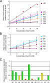
Unstimulated actin assembly activities of wild type and mutant Arp2/3 complexes in the absence of any NPF. A and B, actin (4 μm) was assembled in the presence of variable concentrations of Arp2/3 complex (0-20 nm). Rates of assembly were determined from the slopes of the curves and graphed. C, quantitative comparison of wild type (WT) and mutant Arp2/3 complex activities. The values graphed are the slopes of the titration line fits in A and B, normalized to wild type (arbitrarily set at 1). The bars are color-coded as in Figs. 1 and 3.
FIGURE 5.
FIGURE 5.
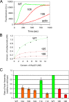
Actin nucleation defects of lethal arc35 mutants. A, actin assembly activities of wild type (WT) and mutant Arp2/3 complexes (15 nm) in the presence of 15 nm yeast WASp/Las17 and 2 μm actin. B, comparison of wild type and mutant Arp2/3 complex activities (expressed as barbed ends generated) at a range of concentrations of Arp2/3 complex (0-15 nm) in the presence of 15 nm WASp. C, comparison of lethal and nonlethal mutant activities. Fold decrease in activity for mutants compared with wild type was determined for a range of concentrations of Arp2/3 complex from the data on lethal (Fig. 5B) and nonlethal (Fig. 3D) mutants. These effects were averaged and are graphed with error bars. All of the bars and assembly curves are color-coded according to mutant growth phenotypes as in Figs. 1, 3, and 4.
FIGURE 6.
FIGURE 6.
Cellular actin organization in arc35 mutants. Wild type (WT) and mutant strains were grown at 25 or 37 °C, fixed, and stained with Alexa 488-phalloidin.
FIGURE 7.
FIGURE 7.
Fluid phase endocytosis defects in arc35 mutants. A, representative images of wild type (WT) and mutant strains compared for uptake of Lucifer yellow dye in the vacuole at 25 °C. B, quantification of defects. The data are averaged from two separate experiments (n > 200 cells for each strain).

References

    1. Goley, E. D., and Welch, M. D. (2006) Nat. Rev. Mol. Cell. Biol. 7 713-726 - PubMed
    1. Machesky, L. M., Atkinson, S. J., Ampe, C., Vandekerckhove, J., and Pollard, T. D. (1994) J. Cell Biol. 127 107-115 - PMC - PubMed
    1. Robinson, R. C., Turbedsky, K., Kaiser, D. A., Marchand, J. B., Higgs, H. N., Choe, S., and Pollard, T. D. (2001) Science 294 1679-1684 - PubMed
    1. Welch, M. D., and Mullins, R. D. (2002) Annu. Rev. Cell Dev. Biol. 18 247-288 - PubMed
    1. Goley, E. D., Rodenbusch, S. E., Martin, A. C., and Welch, M. D. (2004) Mol. Cell 16 269-279 - PubMed

Publication types