. 2008 May 30;283(22):15185-92.
doi: 10.1074/jbc.M802071200. Epub 2008 Apr 1.

Appearance and propagation of polyglutamine-based amyloids in yeast: tyrosine residues enable polymer fragmentation

Affiliations

Appearance and propagation of polyglutamine-based amyloids in yeast: tyrosine residues enable polymer fragmentation

Ilya M Alexandrov et al. J Biol Chem. .

Abstract

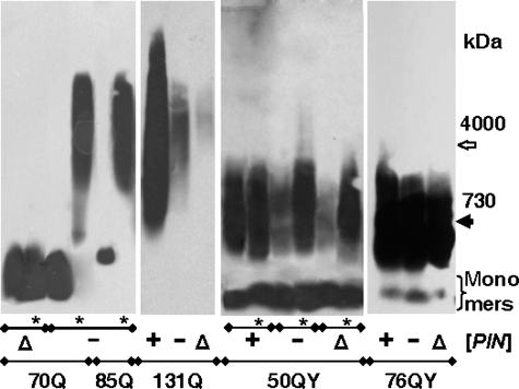
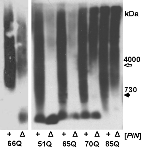
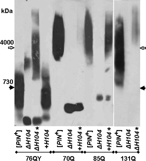
In yeast, fragmentation of amyloid polymers by the Hsp104 chaperone allows them to propagate as prions. The prion-forming domain of the yeast Sup35 protein is rich in glutamine, asparagine, tyrosine, and glycine residues, which may define its prion properties. Long polyglutamine stretches can also drive amyloid polymerization in yeast, but these polymers are unable to propagate because of poor fragmentation and exist through constant seeding with the Rnq1 prion polymers. We proposed that fragmentation of polyglutamine amyloids may be improved by incorporation of hydrophobic amino acid residues into polyglutamine stretches. To investigate this, we constructed sets of polyglutamine with or without tyrosine stretches fused to the non-prion domains of Sup35. Polymerization of these chimeras started rapidly, and its efficiency increased with stretch size. Polymerization of proteins with polyglutamine stretches shorter than 70 residues required Rnq1 prion seeds. Proteins with longer stretches polymerized independently of Rnq1 and thus could propagate. The presence of tyrosines within polyglutamine stretches dramatically enhanced polymer fragmentation and allowed polymer propagation in the absence of Rnq1 and, in some cases, of Hsp104.


Figures

FIGURE 1.
FIGURE 1.
Polymerization of polyQ/QY proteins in [PIN+] cells. A, schematic representation of the polyQ/QY-Sup35MC proteins. The amino acid numbering of native Sup35 is given in parentheses. M, middle domain; C, C-terminal domain. In B–D, the indicated polyQ/QY proteins were expressed in [PIN+] cells instead of endogenous Sup35. B, cell phenotypes on YPD-red medium and synthetic medium lacking adenine (9th day of growth). Sup35MC served as the control. In C and D, cells were grown in YPD medium, and cell lysates were analyzed by electrophoresis. Gel blots were stained with antibody to Sup35M, which reveals polyQ/QY fusion proteins. C, monomer analysis by SDS-PAGE. The samples were not boiled to show the levels of only monomeric polyQ/QY proteins. D, polymer analysis by SDD-AGE. The molecular mass standards titin (4000 kDa) and nebulin (700 kDa) are indicated.
FIGURE 2.
FIGURE 2.
Dependence of polymer appearance on the [PIN] status. Plasmids encoding the 70Q, 85Q, 131Q, 50QY, and 76QY proteins were introduced into the [PIN+], [pin-], and Δrnq1 cells, and transformants were grown for 30 or 100 (*) generations. Cell lysates were analyzed by SDD-AGE with blot immunostaining for Sup35M. Molecular mass standards are indicated in kilodaltons.
FIGURE 3.
FIGURE 3.
Effects of the RNQ1 deletion on propagation of polyQ polymers. The indicated polyQ proteins were expressed in [PIN+] cells (+) and yielded polymers. Then, the RNQ1 gene was disrupted in these cells (Δ). Cell lysates were analyzed by SDD-AGE with blot immunostaining for Sup35M.
FIGURE 4.
FIGURE 4.
Effects of the HSP104 deletion. [PIN+] cells were transformed with plasmids encoding the 76QY, 70Q, 85Q, and 131Q proteins. In these cells, HSP104 was disrupted (ΔH104) and then reintroduced into a centromeric plasmid (+H104). Cell lysates were prepared at 30 or 100 (*) generations after transformation and analyzed by SDD-AGE with blot immunostaining for Sup35M. Molecular mass standards are indicated.
FIGURE 5.
FIGURE 5.
Effects of GdnHCl. A, yeast cells, either wild-type or disrupted for HSP104H104), producing 70Q and 76QY in a polymer form were grown in YPD medium containing 3 mm GdnHCl for the indicated number of generations, and cells with 76QY were then grown for 2 h in the absence of GdnHCl. B, 70Q and 85Q polymers were eliminated in the Δrnq1 strain by growing single cells into colonies in the presence of 3 mm GdnHCl. Cell lysates were analyzed by SDD-AGE with blot staining for Sup35M. The agarose gel concentration was 1.2% for the cells with 70Q and 76QYΔH104 (A) and 1.8% in other cases. Molecular mass standards are indicated.

Similar articles

Cited by

References

    1. Chiti, F., and Dobson, C. M. (2006) Annu. Rev. Biochem. 75 333-366 - PubMed
    1. Prusiner, S. B., Scott, M. R., DeArmond, S. J., and Cohen, F. E. (1998) Cell 93 337-348 - PubMed
    1. Wickner, R. B. (1994) Science 264 566-569 - PubMed
    1. Derkatch, I. L., Bradley, M. E., Zhou, P., Chernoff, Y. O., and Liebman, S. W. (1997) Genetics 147 507-519 - PMC - PubMed
    1. Paushkin, S. V., Kushnirov, V. V., Smirnov, V. N., and Ter-Avanesyan, M. D. (1996) EMBO J. 15 3127-3134 - PMC - PubMed

Publication types

MeSH terms

LinkOut - more resources