. 2008 Jun 6;283(23):15799-806.
doi: 10.1074/jbc.M801553200. Epub 2008 Apr 1.

Dopamine D1 receptor-induced signaling through TrkB receptors in striatal neurons

Affiliations

Dopamine D1 receptor-induced signaling through TrkB receptors in striatal neurons

Yuriko Iwakura et al. J Biol Chem. .

Abstract

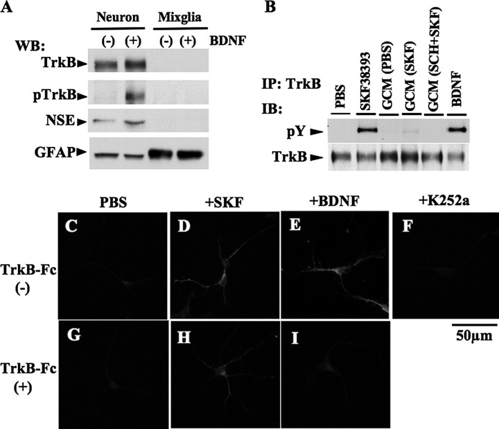
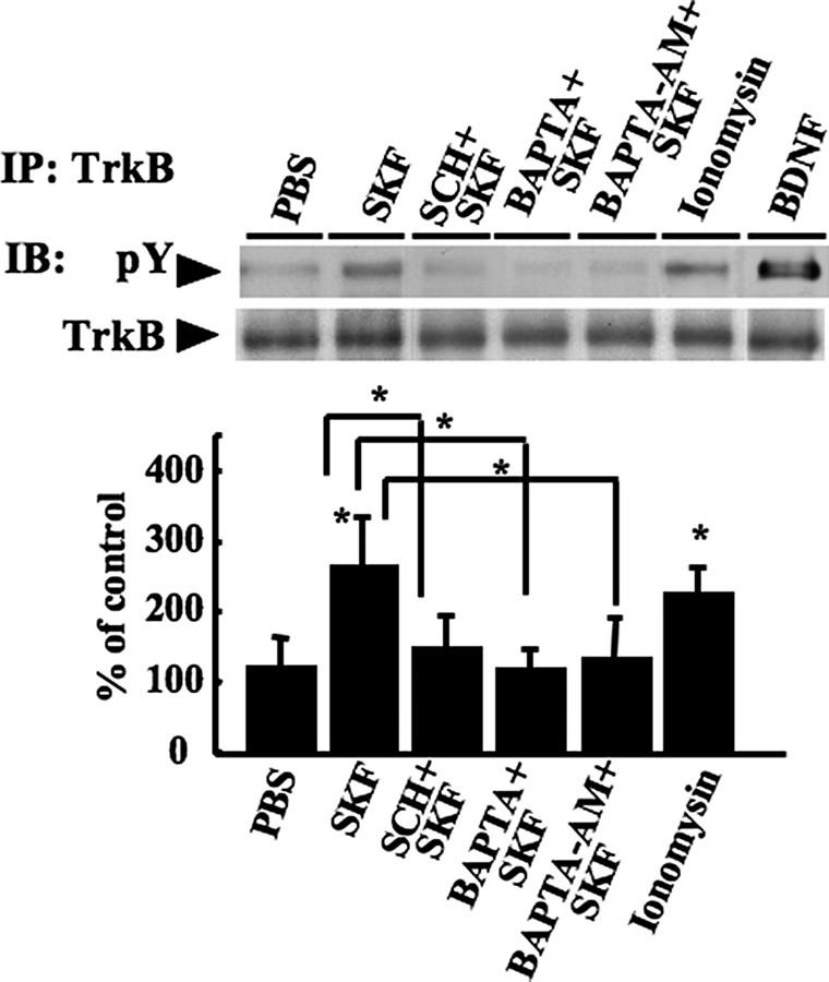
In addition to its role as a neurotransmitter, dopamine can stimulate neurite outgrowth and morphological effects upon primary neurons. To investigate the signal transduction mechanisms used by dopamine in developing striatal neurons, we focused upon the effects of activating the dopamine D1 receptor. Using the D1 receptor agonist SKF38393, we found that Trk neurotrophin receptors were activated in embryonic day 18 striatal neurons. K-252a, a Trk tyrosine kinase inhibitor, and a dopamine D1 receptor antagonist could block the effects of SKF38393. The increase in TrkB phosphorylation was not the result of increased neurotrophin production. Induction of TrkB activity by SKF38393 was accompanied by the phosphorylation of several Trk signaling proteins, including phospholipase Cgamma, Akt, and MAPK. Biotinylation experiments followed by immunostaining by phospho-TrkB-specific antibodies indicated that the mechanism involved increased TrkB surface expression by dopamine D1 receptor activation. This increase in cell surface TrkB expression was dependent upon an increase in intracellular Ca(2+). These results indicate that stimulation of dopamine D1 receptors can be coupled to the neurotrophin receptor signaling to mediate the effects of dopamine upon striatal neurons.


Figures

FIGURE 1.
FIGURE 1.
Trk receptors phosphorylation by D1 receptor agonist in rat striatal cultures. Neuronal striatal cultures were treated with control (PBS), SKF38393 (SKF;1 μm, 10–180 min), quinpirole (QUN;1 μm, 180min), and BDNF (5 ng/ml, 3 min) at 37 °C on DIV6. Some dishes were preincubated with K-252a (K-252a+SKF) or SCH23390 (SCH+SKF) prior to SKF38393 incubation. A, after immunoprecipitation (IP) with Trk antibody, the cell lysates were subjected to SDS-PAGE and Western blotting (WB) for phosphotyrosine (pY) and Trk. Typical immunoblots (IB) are shown. The protein levels were determined from independent wells (n = 4). *, p < 0.05. The bars indicate S.D. B, some of cell lysates for immunoprecipitation were also subjected to Western blotting. Representative immunoblots are shown. Note that treatment with SKF38393 increased the immunoreactivity of growth-associated protein 43 (a growth cone marker) in this culture (data not shown).
FIGURE 2.
FIGURE 2.
Immunoreactivity of phospho-TrkB receptor induced by D1 receptor agonist in rat striatal neurons. A–N, striatal cultures were treated with control (PBS), SKF38393 (SKF), or BDNF at 37 °C on DIV5. Some dishes were preincubated with K-252a or SCH23390 (SCH) prior to SKF38393 incubation. The cultures were fixed and immunostained with phospho-TrkB antibody (red) and dopamine D1 receptor antibody (green). Note that phospho-TrkB immunoreactivity was detected in 87 ± 11% of D1 receptor positive neurons (data not shown). O, specificity of phospho-TrkB antibody was measured by using HEK293-TrkB cells. To assess specificity of phospho-TrkB antibody, cell lysates (30 μg/each) obtained from HEK293-TrkB cells treated with control (PBS) or BDNF (10 ng/ml) for 5 min were probed with phospho-TrkB antibody as well as the antibody that was preincubated with either a phosphorylated or nonphosphorylated TrkB peptide competitor. WB, Western blotting.
FIGURE 3.
FIGURE 3.
Phospho-TrkB level regulated in the striatum of the rats administrated with D1 receptor agonist and antagonist. Littermate rats were injected with control vehicle (PBS; 0.25% ascorbic acid-saline), SKF38393 (A and B, SKF; 1 mg/kg in 0.25% ascorbic acid-saline) and SCH23390 (B, SCH;1 mg/kg in 0.25% ascorbic acid-saline) on postnatal day 4. A, the effects of D1 receptor stimulation at several time points on striatum and frontal cortex were estimated by determining phospho-TrkB. Duplicate samples are displayed. B, the effects of D1 receptor activation and inhibition after 3 h of administration on striatum were estimated by determining phospho-TrkB. Triplicate samples are displayed. Representative immunoblots (IB) are shown. Quantifications of phospho-TrkB immunoreactivity in striatum by NIH Image are shown. Note that there was no significant reduction of the neuron (NSE) marker in the striatum in this pharmacological paradigm (data not shown. control: 100 ± 12.9%, SKF38393: 100.7 ± 7.1%, SCH23390: 107.4 ± 14.8%, mean ± S.D.). n = 4; *, p < 0.05. The bars indicate S.D. IP, immunoprecipitation.
FIGURE 4.
FIGURE 4.
Plasmid transfection to HEK293-TrkB cells. A, HEK293-TrkB cells transfected with mouse D1 receptor expression vector (mD1R) or control vector (mock). After 60 min of incubation with 3H-dopamine (3H-DA)-containing buffer, radioactivity contents were determined from independent wells (n = 4). ***. p < 0.001. The bars indicate S.D. B, mD1R transfected HEK293-TrkB cells were treated with control (PBS), SKF38393 (SKF; 1 μm, 180 min), or BDNF (10 ng/ml, 3 min) at 37 °C. Some dishes were preincubated with K-252a ((+)K252a) or SCH23390 ((+)SCH) prior to SKF38393 incubation. The cell lysates were subjected to SDS-PAGE and Western blotting. Typical immunoblots (IB) are shown.
FIGURE 5.
FIGURE 5.
Effects of glial cells or released factors from culture cells on TrkB transactivation in striatal culture. A, striatal neuronal or glial cultures were treated with control (-) or BDNF (+; 10 ng/ml) for 3 min on DIV6. Cell lysates (30 μg each) were subjected to Western blotting (WB). Representative immunoblots (IB) are shown. B, striatal neuronal cultures were treated with conditioned medium from striatal glial cell culture (GCM) on DIV6. Primary glial cells were treated with control (PBS), SKF38393 (SKF; 1 μm, 3 h), or SCH23390 (1 μm, 1 h) prior to SKF38393 (SCH+SKF). After 3 h of incubation, the media were harvested and used as glial condition media for striatal neuronal culture. Neuronal cells were lysed after the treatment with SKF38393, each glial conditioned medium, or BDNF, and then cell samples were immunoprecipitated (IP) with anti-TrkB antibody. Representative immunoblots are shown. C–I, striatal cultures were treated with control (PBS), SKF38393 (+SKF), K-252a and BDNF on DIV6. Some of the cultures were preincubated with TrkB-Fc (10 μg/ml, for 20 min) in 37 °C prior to control (G), SKF38393 (H), or BDNF (I). The cultures were fixed and immunostained with anti-phospho-TrkB antibody.
FIGURE 6.
FIGURE 6.
Intracellular Ca2+ regulation is involved in the TrkB transactivation by D1 receptor agonist. Striatal cultures were treated with control, SKF38393 (SKF), ionomycin (10 μm), and BDNF on DIV6. Some of the cultures were preincubated with SCH23390 (SCH+SKF), BAPTA (BAPTA+SKF; 10 μm for 30 min), or BAPTA-AM (BAPTA-AM+SKF; 10 μm for 30 min) in 37 °C prior to SKF38393. After immunoprecipitation (IP), the cell lysates were subjected to SDS-PAGE and Western blotting for phosphotyrosine (pY) and TrkB. Duplicate samples are displayed. Typical immunoblots (IB) are shown. The protein levels were determined from independent wells (n = 4). *, p < 0.05. The bars indicate S.D.
FIGURE 7.
FIGURE 7.
Surface TrkB expression regulated by D1 receptor agonist. Striatal cultures were treated with control vehicle (PBS), SKF38393 (SKF), and BDNF on DIV6. Some of the cultures were preincubated with SCH23390 (A, SCH+SKF), K-252a (A), BAPTA (B, BAPTA+SKF), or BAPTA-AM (B, BAPTA-AM+SKF) in 37 °C prior to SKF38393. The cultures were incubated with biotin-PBS for 30 min on ice and then lysed with radioimmune precipitation assay buffer. Half of biotinylated proteins were precipitated by avidin-conjugated beads and then subjected to SDS-PAGE and Western blotting for TrkB. The other samples were used for immunoprecipitation (IP) with TrkB. Duplicate samples are displayed. Typical immunoblots (IB) are shown. Protein levels were determined from independent wells (n = 4). Quantifications of surface biotinylated TrkB levels by NIH Image are shown. *, p < 0.05. The bars indicate S.D. CH, striatal cultures were treated with control (PBS) and SKF38393 on DIV6. Some of the cultures were preincubated with SCH23390 (SCH+SKF), BAPTA (BAPTA+SKF), or BAPTA-AM (AM+SKF) in 37 °C prior to SKF38393. The cultures were fixed and immunostained with rabbit control IgG (C) or TrkB (N terminus) antibody (D–H) without detergent. Representative pictures are provided.

References

    1. Kalivas, P. W. (1993) Brain Res. 18 75-113 - PubMed
    1. Missale, C., Nash, S. R., Robinson, S. W., Jaber, M., and Caron, M. G. (1998) Physiol. Rev. 78 189-225 - PubMed
    1. Bustos, G., Abarca, J., Campusano, J., Bustos, V., Noriega, V., and Aliaga, E. (2004) Brain Res. 47 126-144 - PubMed
    1. Xu, M., Hu, X. T., Cooper, D. C., Moratalla, R., Graybiel, A. M., White, F. J., and Tonegawa, S. (1994) Cell 79 945-955 - PubMed
    1. Centonze, D., Grande, C., Saulle, E., Martin, A. B., Gubellini, P., Pavon, N., Pisani, A., Bernardi, G., Moratalla, R., and Calabresi, P. (2003) J. Nuerosci. 23 8506-8512 - PMC - PubMed

Publication types

MeSH terms