

Noninvasive quantification of human nucleus pulposus pressure with use of T1rho-weighted magnetic resonance imaging
- PMID: 18381318
- PMCID: PMC2657301
- DOI: 10.2106/JBJS.G.00667
Noninvasive quantification of human nucleus pulposus pressure with use of T1rho-weighted magnetic resonance imaging
Abstract
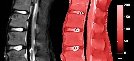
Background: Early diagnosis is a challenge in the treatment of degenerative disc disease. A noninvasive biomarker detecting functional mechanics of the disc is needed. T1rho-weighted imaging, a spin-lock magnetic resonance imaging technique, has shown promise for meeting this need in in vivo studies demonstrating the clinical feasibility of evaluating both intervertebral discs and articular cartilage. The objectives of the present study were (1) to quantitatively determine the relationship between T1rho relaxation time and measures of nucleus pulposus mechanics, and (2) to evaluate whether the quantitative relationship of T1rho relaxation time with the degenerative grade and glycosaminoglycan content extend to more severe degeneration. It was hypothesized that the isometric swelling pressure and compressive modulus would be directly correlated with the T1rho relaxation time and the apparent permeability would be inversely correlated with the T1rho relaxation time.
Methods: Eight cadaver human lumbar spines were imaged to measure T1rho relaxation times. The nucleus pulposus tissue from the L1 disc through the S1 disc was tested in confined compression to determine the swelling pressure, compressive modulus, and permeability. The glycosaminoglycan and water contents were measured in adjacent tissue. Linear regression analyses were performed to examine the correlation between the T1rho relaxation time and the other measured variables. Mechanical properties and biochemical content were evaluated for differences associated with degeneration.
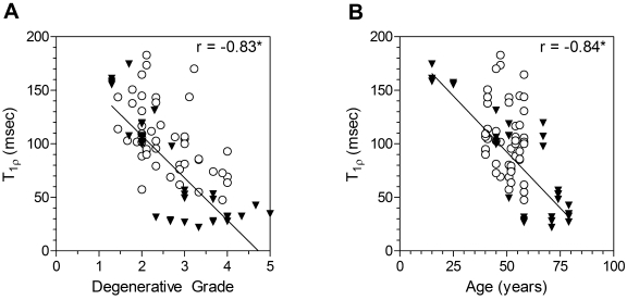
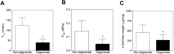
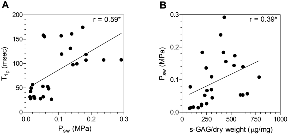
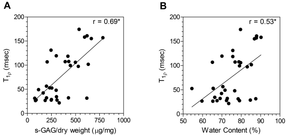
Results: A positive linear correlation was observed between the T1rho relaxation time on the images of the nucleus pulposus and the swelling pressure (r = 0.59), glycosaminoglycan content per dry weight (r = 0.69), glycosaminoglycan per wet weight (r = 0.49), and water content (r = 0.53). No significant correlations were observed between the T1rho relaxation time and the modulus or permeability. Similarly, the T1rho relaxation time, swelling pressure, glycosaminoglycan content per dry weight, and water content were significantly altered with degeneration, whereas the modulus and permeability were not.
Conclusions: T1rho-weighted magnetic resonance imaging has a strong potential as a quantitative biomarker of the mechanical function of the nucleus pulposus and of disc degeneration.
Figures
References
-
- Johannessen W, Vresilovic EJ, Wright AC, Elliott DM. Intervertebral disc mechanics are restored following cyclic loading and unloaded recovery. Ann Biomed Eng. 2004;32:70-6. - PubMed
-
- Guerin HAL, Elliott DM. Structure and properties of soft tissues in the spine. In: Kurtz SM, Edidin AA, editors. Spine technology handbook. San Diego: Elsevier Academic Press; 2006. p 35-62.
-
- Urban JP, Maroudas A. The chemistry of the intervertebral disc in relation to its physiological function and requirements. Clin Rheum Dis. 1980;6:51-76.
-
- Buckwalter JA. Aging and degeneration of the human intervertebral disc. Spine. 1995;20:1307-14. - PubMed
-
- Pearce RH, Grimmer BJ, Adams ME. Degeneration and the chemical composition of the human lumbar intervertebral disc. J Orthop Res. 1987;5:198-205. - PubMed
MeSH terms
Substances
LinkOut - more resources
Full Text Sources
Other Literature Sources
Medical
