. 2008 Apr 1;68(7):2286-91.
doi: 10.1158/0008-5472.CAN-07-1349.

Detection of early prostate cancer using a hepsin-targeted imaging agent

Affiliations

Detection of early prostate cancer using a hepsin-targeted imaging agent

Kimberly A Kelly et al. Cancer Res. .

Abstract

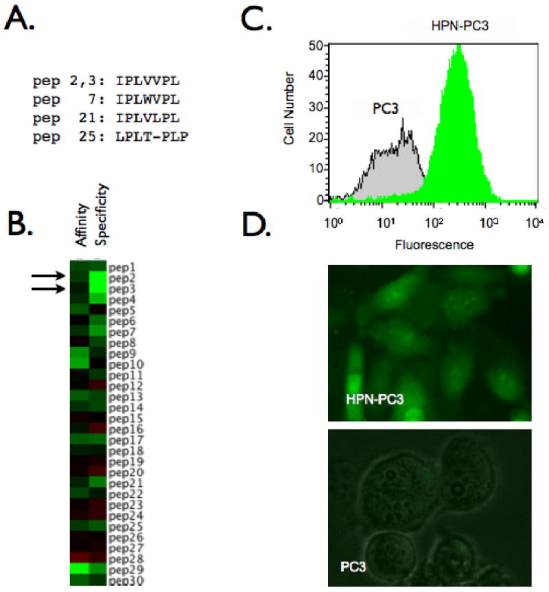
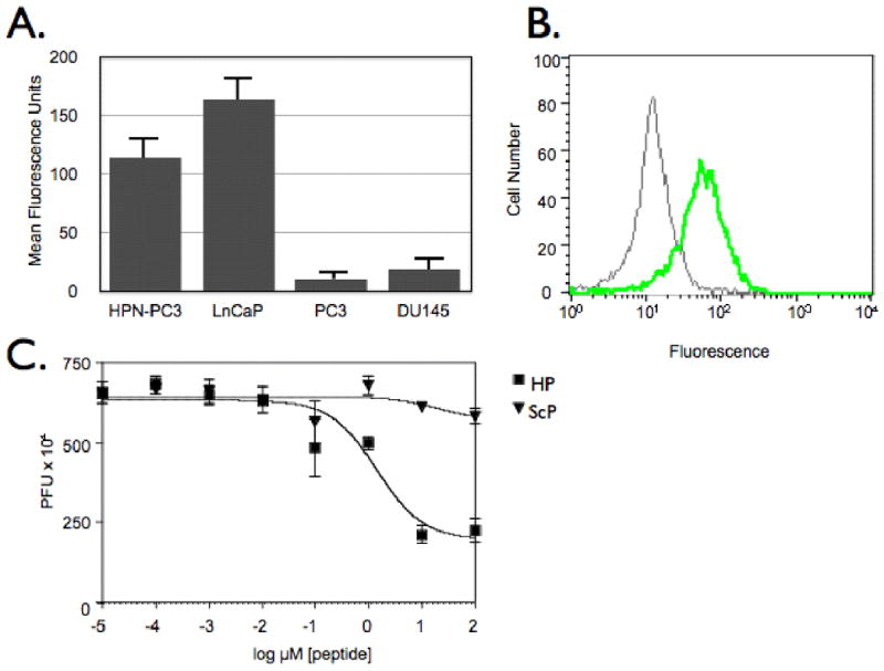
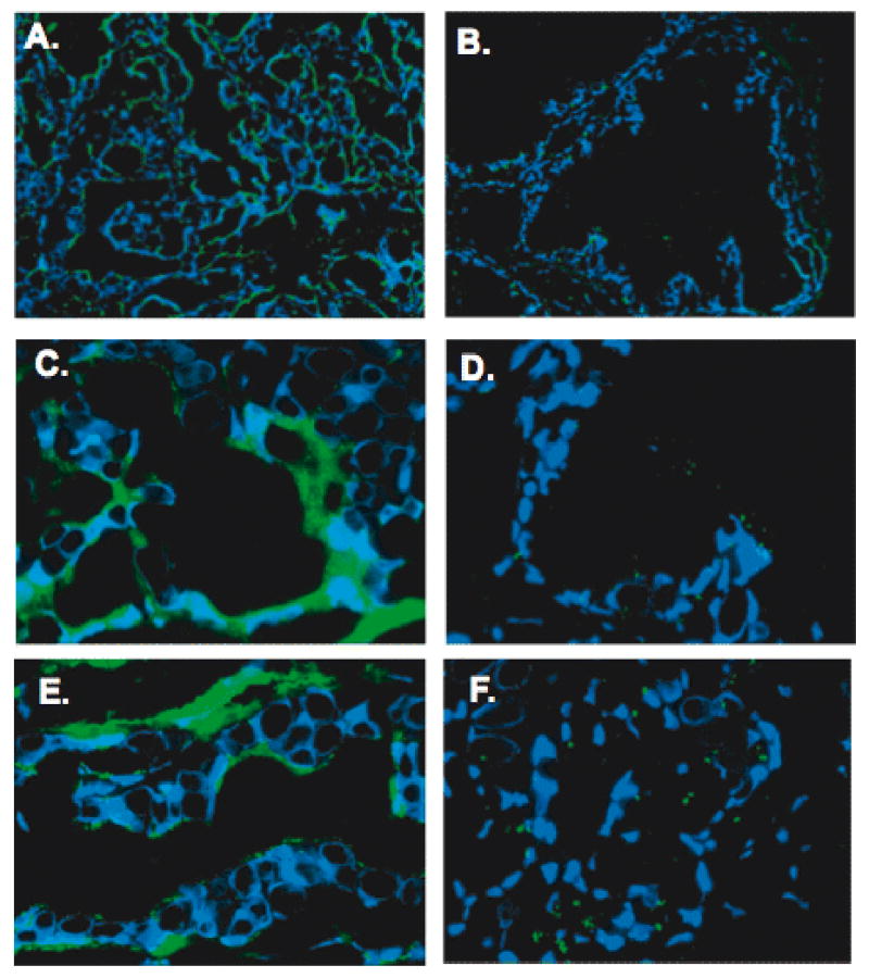
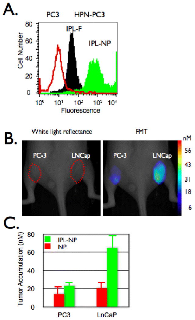
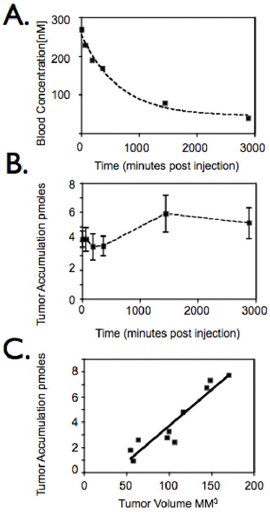
Early detection and diagnosis of prostate cancer is key to designing effective treatment strategies. Microarrays have resulted in the discovery of hepsin (HPN) as a biomarker for detection of prostate cancer. In this study, we explore the development of HPN imaging probes for detection of prostate cancer. We used phage display to isolate HPN binding peptides with 190 + 2.2 nmol/L affinity in monomeric form and high specificity. The identified peptides were able to detect human prostate cancer on tissue microarrays and in cell-based assays. HPN-targeted imaging agents were synthesized by conjugating multiple peptides to fluorescent nanoparticles to further improve avidity through multivalency and to improve pharmacokinetics. When injected into mouse xenograft models, HPN-targeted nanoparticles bound specifically to HPN-expressing LNCaP xenografts compared with non-HPN-expressing PC3 xenografts. HPN imaging may provide a new method for detection of prostate cancer.


Figures

Figure 1
Figure 1
Phage display screening produces a peptide specific for HPN. A. Consensus family identified through selection B. Heatmap depicting affinity and specificity of individual phage clones. Green: high value, black: average value, red: low value. C. FACs analysis demonstrating specificity of IPL-• for HPN-PC3 cells. D. Fluorescence microscope images of IPL-• binding to HPN-PC3 cells but not parental PC3 cells.
Figure 2
Figure 2
Characterization of IPL-F binding to HPN-expressing cells. A. IPL-F discriminates between HPN expressing prostate cancer cell lines HPN-PC3 and LNCaP and non-expressing cell lines PC3 and DU145. B. IPL-F binds to HPN. Anti-HPN antibody competes for IPL-F binding to HPN-PC3 cells. Green Histogram: HPN-PC3 cells incubated with IPL-F, Gray Histogram: HPN-PC3 cells incubated with anti-HPN antibody and IPL-F. C. Competition assay. IPL-• were competed from HPN-PC3 cells with increasing concentrations of IPL-F whereas ScP was unable to compete for binding at assayed concentrations of peptide
Figure 3
Figure 3
HPN targeted peptides and nanoparticles are able to identify human prostate cancer. IPL-F or IPL-NP was incubated with tissue microarrays consisting of 6 normal and 5 cancer human radical prostatectomy specimens. Results shown are representative images. Panels A. and C show the binding of IPL-F/IPL-NP to cancer glands and panels B. and D. show the binding to benign glands (Original magnification of A and B, 20×; Original magnification of C and D, 60×). Note staining in the tumor cells and the absence/weak staining in benign tissues and stroma.
Figure 4
Figure 4
In vivo imaging of prostate cancer. A. HPN-PC3 (black and green histograms) or PC3 (red histogram) cells were incubated with IPL-F (black histogram) or IPL-NP (green histogram) then analyzed via flow cytometry. B and C. Mice bearing tumors derived from PC-3 (left flank) or LNCaP (right flank) were co-injected with IPL-NP (green bars) and NP (red bars) then (B) imaged and (C) accumulation quantified via FMT 24 hours post injection.
Figure 5
Figure 5
In vivo characterization of IPL-NP. A. Mice bearing tumors derived from LNCaP cells were injected with IPL-NP (20 mg/kg Fe) then blood analyzed for agent presence at indicated time points post injection. B. Time course of IPL-NP accumulation into LNCaP derived tumors. Mice bearing LNCaP derived tumors were injected with IPL-NP (20 mg/kg Fe) then imaged via FMT at 0,1,3,6,24, and 48 hours post injection. C. Mice bearing tumors of different volumes derived from LNCaP cells were injected with identical doses of IPL-NP (20 mg/kg Fe) then imaged via FMT.

References

    1. Jemal A, Siegel R, Ward E, Murray T, Xu J, Thun MJ. Cancer statistics, 2007. CA Cancer J Clin. 2007;57:43–66. - PubMed
    1. Bill-Axelson A, Holmberg L, Ruutu M, et al. Radical prostatectomy versus watchful waiting in early prostate cancer. N Engl J Med. 2005;352:1977–84. - PubMed
    1. D'Amico AV, Whittington R, Malkowicz SB, et al. Biochemical outcome after radical prostatectomy, external beam radiation therapy, or interstitial radiation therapy for clinically localized prostate cancer. JAMA. 1998;280:969–74. - PubMed
    1. Sakr WA, Haas GP, Cassin BF, Pontes JE, Crissman JD. The frequency of carcinoma and intraepithelial neoplasia of the prostate in young male patients. J Urol. 1993;150:379–85. - PubMed
    1. Thompson IM, Pauler DK, Goodman PJ, et al. Prevalence of prostate cancer among men with a prostate-specific antigen level < or =4.0 ng per milliliter. N Engl J Med. 2004;350:2239–46. - PubMed

Publication types