

Inhibition of SNAP25 expression by HIV-1 Tat involves the activity of mir-128a
- PMID: 18381601
- PMCID: PMC2662126
- DOI: 10.1002/jcp.21452
Inhibition of SNAP25 expression by HIV-1 Tat involves the activity of mir-128a
Abstract
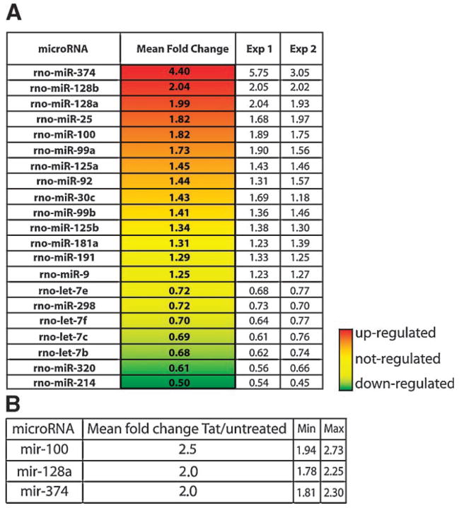
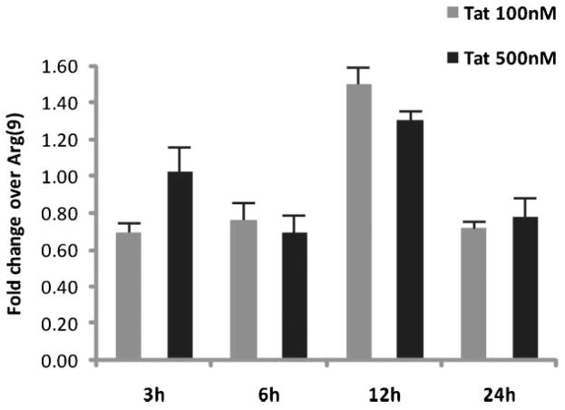
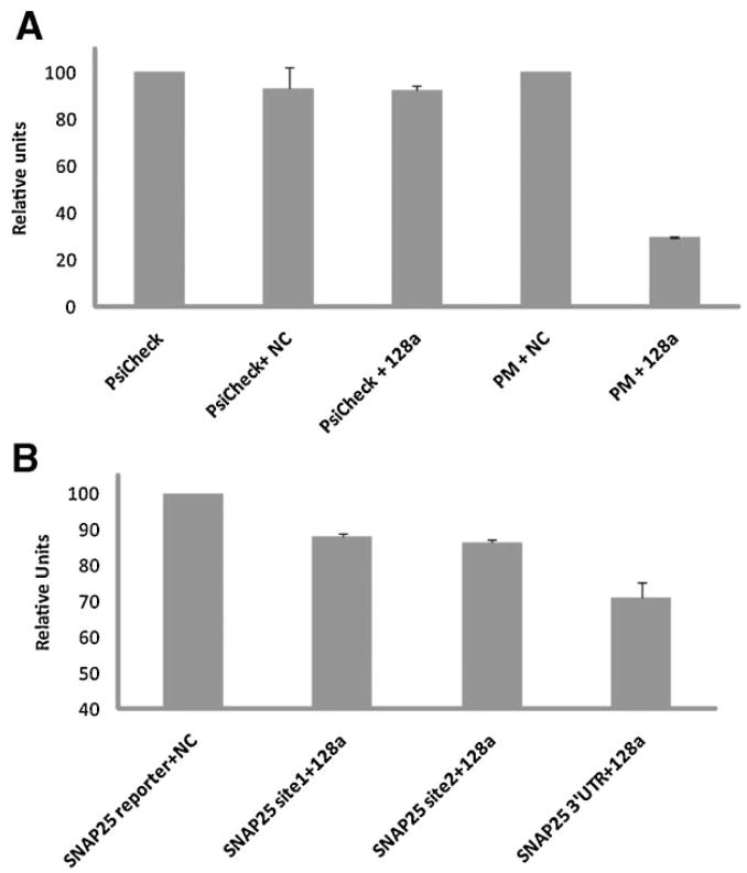
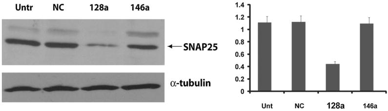
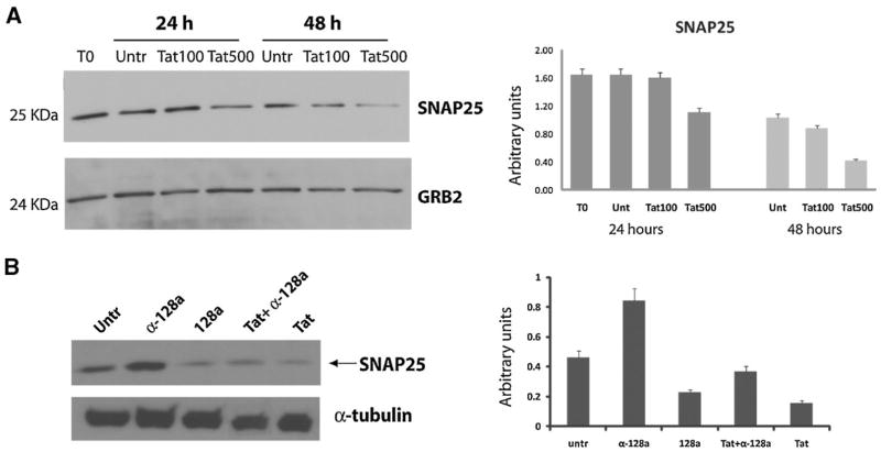
MicroRNAs (miRs) are short endogenous RNAs that regulate gene expression by incomplete pairing with messenger RNAs. An increasing number of studies show that mammalian microRNAs play fundamental roles in various aspects of cellular function including differentiation, proliferation, and cell death. Recent findings demonstrating the presence of microRNAs in mature neuronal dendrites suggest their possible involvement in controlling local protein translation and synaptic function. HIV-1 Encephalopathy (HIVE) is a manifestation of HIV-1 infection that often results in neuronal damage and dysfunction. While neurons are rarely, if ever, infected by HIV-1, they are exposed to cytotoxic viral and cellular factors including the HIV-1 transactivating factor Tat. In this study, we show that Tat deregulates expression levels of selected microRNAs, including the neuronal mir-128, in primary cortical neurons. We further show that mir-128a inhibits expression of the pre-synaptic protein SNAP25, whereas the anti-mir-128a partially restores Tat/mir-128a-induced downregulation of SNAP25 expression. Altogether, our data provide a novel mechanism by which HIV-Tat perturbs neuronal activity.
Figures
Similar articles
-
HIV-1 Tat protein promotes neuronal dysfunction through disruption of microRNAs.J Biol Chem. 2011 Nov 25;286(47):41125-34. doi: 10.1074/jbc.M111.268466. Epub 2011 Sep 28. J Biol Chem. 2011. PMID: 21956116 Free PMC article.
-
Novel insights into role of miR-320a-VDAC1 axis in astrocyte-mediated neuronal damage in neuroAIDS.Glia. 2017 Feb;65(2):250-263. doi: 10.1002/glia.23089. Epub 2016 Oct 20. Glia. 2017. PMID: 27761954
-
HIV Tat induces expression of ICAM-1 in HUVECs: implications for miR-221/-222 in HIV-associated cardiomyopathy.PLoS One. 2013;8(3):e60170. doi: 10.1371/journal.pone.0060170. Epub 2013 Mar 28. PLoS One. 2013. PMID: 23555914 Free PMC article.
-
Involvement of miR-196a in HIV-associated neurocognitive disorders.Apoptosis. 2014 Aug;19(8):1202-14. doi: 10.1007/s10495-014-1003-2. Apoptosis. 2014. PMID: 24872081
-
Role of Tat-interacting protein of 110 kDa and microRNAs in the regulation of hematopoiesis.Curr Opin Hematol. 2016 Jul;23(4):325-30. doi: 10.1097/MOH.0000000000000246. Curr Opin Hematol. 2016. PMID: 27071021 Review.
Cited by
-
Role of Tat protein in HIV neuropathogenesis.Neurotox Res. 2009 Oct;16(3):205-20. doi: 10.1007/s12640-009-9047-8. Epub 2009 Mar 21. Neurotox Res. 2009. PMID: 19526283 Review.
-
Glia-to-neuron transfer of miRNAs via extracellular vesicles: a new mechanism underlying inflammation-induced synaptic alterations.Acta Neuropathol. 2018 Apr;135(4):529-550. doi: 10.1007/s00401-017-1803-x. Epub 2018 Jan 4. Acta Neuropathol. 2018. PMID: 29302779 Free PMC article.
-
Genetic, transcriptomic, and epigenetic studies of HIV-associated neurocognitive disorder.J Acquir Immune Defic Syndr. 2014 Apr 1;65(4):481-503. doi: 10.1097/QAI.0000000000000069. J Acquir Immune Defic Syndr. 2014. PMID: 24583618 Free PMC article. Review.
-
A miRNA Signature for Cognitive Deficits and Alcohol Use Disorder in Persons Living with HIV/AIDS.Front Mol Neurosci. 2017 Nov 15;10:385. doi: 10.3389/fnmol.2017.00385. eCollection 2017. Front Mol Neurosci. 2017. PMID: 29187813 Free PMC article.
-
Chronic, Low-Dose Methamphetamine Reveals Sexual Dimorphism of Memory Performance, Histopathology, and Gene Expression Affected by HIV-1 Tat Protein in a Transgenic Model of NeuroHIV.Viruses. 2025 Feb 28;17(3):361. doi: 10.3390/v17030361. Viruses. 2025. PMID: 40143289 Free PMC article.
References
-
- Bennasser Y, Le SY, Benkirane M, Jeang KT. Evidence that HIV-1 encodes an siRNA and a suppressor of RNA silencing. Immunity. 2005;22:607–619. - PubMed
-
- Burre J, Volknandt W. The synaptic vesicle proteome. J Neurochem. 2007;101:1448–1462. - PubMed
-
- Cao X, Yeo G, Muotri AR, Kuwabara T, Gage FH. Noncoding Rnas in the mammalian central nervous system. Annu Rev Neurosci. 2006;29:77–103. - PubMed
Publication types
MeSH terms
Substances
Grants and funding
LinkOut - more resources
Full Text Sources
