

Dynorphin and stress-related peptides in rat locus coeruleus: contribution of amygdalar efferents
- PMID: 18381633
- PMCID: PMC3277290
- DOI: 10.1002/cne.21683
Dynorphin and stress-related peptides in rat locus coeruleus: contribution of amygdalar efferents
Abstract
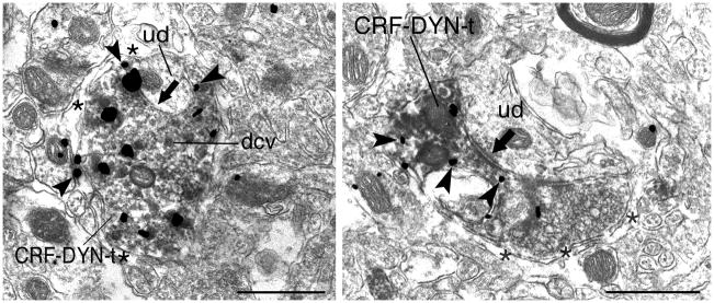
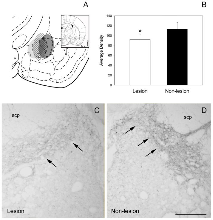
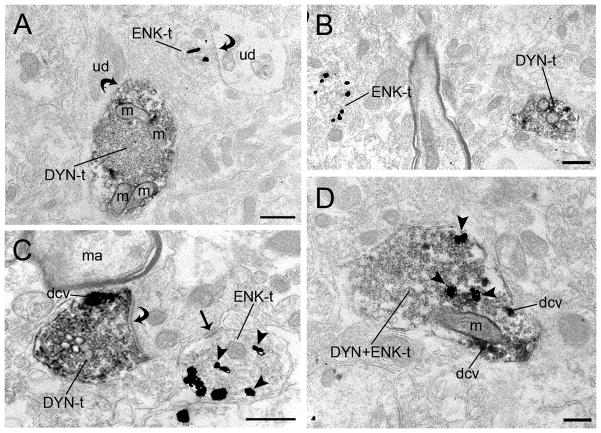
The interaction between the stress axis and endogenous opioid systems has gained substantial attention, because it is increasingly recognized that stress alters individual sensitivity to opiates. One site at which opiates and stress substrates may interact to have global effects on behavior is within the locus coeruleus (LC). We have previously described interactions of several opioid peptides [e.g., proopiomelanocortin, enkephalin (ENK)] with the stress-related peptide corticotropin-releasing factor (CRF) in the LC. To examine further the interactions among dynorphin (DYN), ENK, and CRF in the LC, sections were processed for detection of DYN and CRF or DYN and ENK in rat brain. DYN- and CRF-containing axon terminals overlapped noradrenergic dendrites in this region. Dual immunoelectron microscopy showed coexistence of DYN and CRF; 35% of axon terminals containing DYN were also immunoreactive for CRF. In contrast, few axon terminals contained both DYN and ENK. A potential DYN/CRF afferent is the central nucleus of the amygdala (CeA). Dual in situ hybridization showed that, in CeA neurons, 31% of DYN mRNA-positive cells colocalized with CRF mRNA, whereas 53% of CRF mRNA-containing cells colocalized with DYN mRNA. Finally, to determine whether limbic DYN afferents target the LC, the CeA was electrolytically lesioned. Light-level densitometry of DYN labeling in the LC showed a significant decrease in immunoreactivity on the side of the lesion. Taken together, these data indicate that DYN- and CRF-labeled axon terminals, most likely arising from amygdalar sources, are positioned dually to affect LC function, whereas DYN and ENK function in parallel.
Figures


Similar articles
-
Cellular interactions between axon terminals containing endogenous opioid peptides or corticotropin-releasing factor in the rat locus coeruleus and surrounding dorsal pontine tegmentum.J Comp Neurol. 2003 Nov 24;466(4):445-56. doi: 10.1002/cne.10893. J Comp Neurol. 2003. PMID: 14566941
-
Amygdalar peptidergic circuits regulating noradrenergic locus coeruleus neurons: linking limbic and arousal centers.Exp Neurol. 2011 Jul;230(1):96-105. doi: 10.1016/j.expneurol.2011.04.001. Epub 2011 Apr 16. Exp Neurol. 2011. PMID: 21515261 Free PMC article.
-
Direct targeting of peptidergic amygdalar neurons by noradrenergic afferents: linking stress-integrative circuitry.Brain Struct Funct. 2015 Jan;220(1):541-58. doi: 10.1007/s00429-013-0674-8. Epub 2013 Nov 23. Brain Struct Funct. 2015. PMID: 24271021 Free PMC article.
-
Opposing regulation of the locus coeruleus by corticotropin-releasing factor and opioids. Potential for reciprocal interactions between stress and opioid sensitivity.Psychopharmacology (Berl). 2001 Dec;158(4):331-42. doi: 10.1007/s002130000673. Epub 2001 Mar 14. Psychopharmacology (Berl). 2001. PMID: 11797054 Review.
-
Physiological and neurochemical aspects of corticotropin-releasing factor actions in the brain: the role of the locus coeruleus.Neurochem Res. 1998 Aug;23(8):1039-52. doi: 10.1023/a:1020751817723. Neurochem Res. 1998. PMID: 9704593 Review.
Cited by
-
Protracted withdrawal from ethanol and enhanced responsiveness stress: regulation via the dynorphin/kappa opioid receptor system.Alcohol. 2013 Aug;47(5):359-65. doi: 10.1016/j.alcohol.2013.05.001. Epub 2013 May 31. Alcohol. 2013. PMID: 23731692 Free PMC article.
-
Increased GABAergic Efficacy of Central Amygdala Projections to Neuropeptide S Neurons in the Brainstem During Fear Memory Retrieval.Neuropsychopharmacology. 2015 Nov;40(12):2753-63. doi: 10.1038/npp.2015.125. Epub 2015 May 4. Neuropsychopharmacology. 2015. PMID: 25936641 Free PMC article.
-
Reciprocal midbrain-extended amygdala circuit activity in preclinical models of alcohol use and misuse.Neuropharmacology. 2022 Jan 1;202:108856. doi: 10.1016/j.neuropharm.2021.108856. Epub 2021 Oct 25. Neuropharmacology. 2022. PMID: 34710467 Free PMC article. Review.
-
Co-localization of the cannabinoid type 1 receptor with corticotropin-releasing factor-containing afferents in the noradrenergic nucleus locus coeruleus: implications for the cognitive limb of the stress response.Brain Struct Funct. 2017 Sep;222(7):3007-3023. doi: 10.1007/s00429-017-1381-7. Epub 2017 Mar 2. Brain Struct Funct. 2017. PMID: 28255675 Free PMC article.
-
Coordinate regulation of noradrenergic and serotonergic brain regions by amygdalar neurons.J Chem Neuroanat. 2013 Sep;52:9-19. doi: 10.1016/j.jchemneu.2013.04.003. Epub 2013 May 4. J Chem Neuroanat. 2013. PMID: 23651691 Free PMC article.
References
-
- Abercrombie ED, Jacobs BL. Systemic naloxone administration potentiates locus coeruleus noradrenergic neuronal activity under stressful but not non-stressful conditions. Brain Res. 1988;441:362–366. - PubMed
-
- Akil H, Watson SJ, Young E, Lewis ME, Khachaturian H, Walker JM. Endogenous opioids: biology and function. Annu Rev Neurosci. 1984;7:223–255. - PubMed
-
- Arvidsson U, Riedl M, Chakrabarti S, Vulchanova L, Lee JH, Nakano AH, Lin X, Loh HH, Law PY, Wessendorf MW, Elde R. The kappa-opioid receptor is primarily postsynaptic: combined immunohistochemical localization of the receptor and endogenous opioids. Proc Natl Acad Sci USA. 1995;92:5062–5066. - PMC - PubMed
Publication types
MeSH terms
Substances
Grants and funding
LinkOut - more resources
Full Text Sources
