. 2008 Apr;9(4):329-34.
doi: 10.1631/jzus.B0710546.

Values of endoscopic ultrasonography for diagnosis and treatment of duodenal protruding lesions

Affiliations

Values of endoscopic ultrasonography for diagnosis and treatment of duodenal protruding lesions

Guo-qiang Xu et al. J Zhejiang Univ Sci B. 2008 Apr.

Abstract

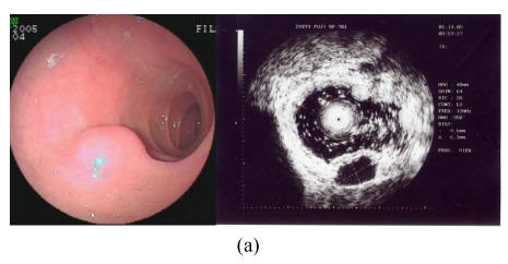
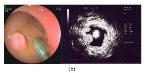
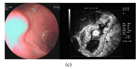
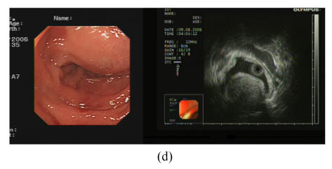
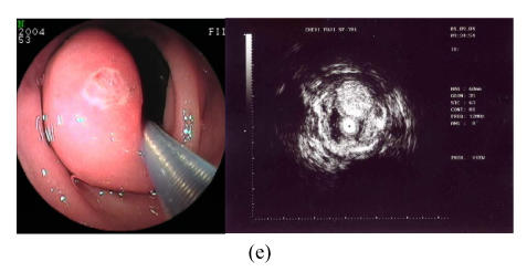
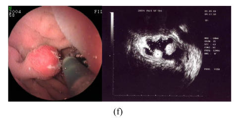
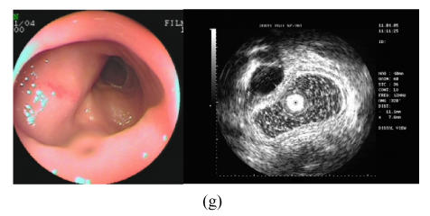
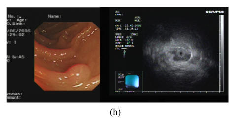
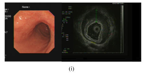
Objective: The diagnoses of patients with duodenal protruding lesions are difficult when using conventional examinations such as computed tomography (CT) and conventional endoscope etc. Thus, we investigated the clinical value of endoscopic ultrasonography (EUS) with miniature ultrasonic probes on the diagnosis and treatment of duodenal protruding lesions.

Methods: Patients with duodenal protruding lesions who were indicated for EUS were examined by EUS with 12 approximately 15 MHz miniature ultrasonic probes and double-cavity electronic endoscope. According to diagnosis of EUS, those patients were indicated for biopsy and treatment received biopsy, endoscopic resection or surgical excision. The postoperative histological results were compared with the preoperative diagnosis of EUS. Those patients without endoscopic resection or surgical excision were periodically followed up with EUS.

Results: A total of 169 patients with duodenal protruding lesions were examined by EUS, of which 40 were diagnosed with cysts, 36 with inflammatory protruding or polyp, 25 with Brunner's gland adenoma, 19 with ectopic pancreas, 17 with gastrointestinal stromal tumor, 12 with extrinsic compression, 12 with minor papilla, 6 with lipoma, 1 with adenocarcinoma and 1 with lymphoma. After EUS examinations, 75 patients received biopsy, endoscopic resection or surgical excision respectively. The postoperative histological results of 70 patients were completely consistent with the preoperative diagnosis of EUS, with 93.33% diagnostic accuracy. The results of follow-up with EUS indicated that duodenal cyst, Brunner's gland adenoma, ectopic pancreas, gastrointestinal stromal tumor and lipoma remained unchanged within 1 approximately 3 years. No related complications occurred among all patients that received EUS examinations.

Conclusion: EUS is an effective and reliable diagnostic method for duodenal protruding lesions.


Figures

Fig. 1
Fig. 1
(a) Duodenal cyst; (b) Duodenal Brunner’s gland adenoma; (c) Duodenal ectopic pancreas; (d) Duodenal stromal tumor; (e) Duodenal lipoma; (f) Duodenal polyp; (g) Duodenal extrinsic compression (cholecyst); (h) Duodenal minor papilla; (i) Duodenal B-cell lymphoma
Fig. 1
Fig. 1
(a) Duodenal cyst; (b) Duodenal Brunner’s gland adenoma; (c) Duodenal ectopic pancreas; (d) Duodenal stromal tumor; (e) Duodenal lipoma; (f) Duodenal polyp; (g) Duodenal extrinsic compression (cholecyst); (h) Duodenal minor papilla; (i) Duodenal B-cell lymphoma
Fig. 1
Fig. 1
(a) Duodenal cyst; (b) Duodenal Brunner’s gland adenoma; (c) Duodenal ectopic pancreas; (d) Duodenal stromal tumor; (e) Duodenal lipoma; (f) Duodenal polyp; (g) Duodenal extrinsic compression (cholecyst); (h) Duodenal minor papilla; (i) Duodenal B-cell lymphoma
Fig. 1
Fig. 1
(a) Duodenal cyst; (b) Duodenal Brunner’s gland adenoma; (c) Duodenal ectopic pancreas; (d) Duodenal stromal tumor; (e) Duodenal lipoma; (f) Duodenal polyp; (g) Duodenal extrinsic compression (cholecyst); (h) Duodenal minor papilla; (i) Duodenal B-cell lymphoma
Fig. 1
Fig. 1
(a) Duodenal cyst; (b) Duodenal Brunner’s gland adenoma; (c) Duodenal ectopic pancreas; (d) Duodenal stromal tumor; (e) Duodenal lipoma; (f) Duodenal polyp; (g) Duodenal extrinsic compression (cholecyst); (h) Duodenal minor papilla; (i) Duodenal B-cell lymphoma
Fig. 1
Fig. 1
(a) Duodenal cyst; (b) Duodenal Brunner’s gland adenoma; (c) Duodenal ectopic pancreas; (d) Duodenal stromal tumor; (e) Duodenal lipoma; (f) Duodenal polyp; (g) Duodenal extrinsic compression (cholecyst); (h) Duodenal minor papilla; (i) Duodenal B-cell lymphoma
Fig. 1
Fig. 1
(a) Duodenal cyst; (b) Duodenal Brunner’s gland adenoma; (c) Duodenal ectopic pancreas; (d) Duodenal stromal tumor; (e) Duodenal lipoma; (f) Duodenal polyp; (g) Duodenal extrinsic compression (cholecyst); (h) Duodenal minor papilla; (i) Duodenal B-cell lymphoma
Fig. 1
Fig. 1
(a) Duodenal cyst; (b) Duodenal Brunner’s gland adenoma; (c) Duodenal ectopic pancreas; (d) Duodenal stromal tumor; (e) Duodenal lipoma; (f) Duodenal polyp; (g) Duodenal extrinsic compression (cholecyst); (h) Duodenal minor papilla; (i) Duodenal B-cell lymphoma
Fig. 1
Fig. 1
(a) Duodenal cyst; (b) Duodenal Brunner’s gland adenoma; (c) Duodenal ectopic pancreas; (d) Duodenal stromal tumor; (e) Duodenal lipoma; (f) Duodenal polyp; (g) Duodenal extrinsic compression (cholecyst); (h) Duodenal minor papilla; (i) Duodenal B-cell lymphoma

Similar articles

Cited by

References

    1. Arguello L. Endoscopic ultrasonography in submucosal lesions and extrinsic compressions of the gastrointestinal tract. Minerva Med. 2007;98(4):389–393. - PubMed
    1. Brand B, Oesterhelweg L, Binmoeller KF, Srirann PV, Bohnacker S, Seewald S, Weerth AD, Soehendra N. Impact of endoscopic ultrasound for evaluation of submucosal lesions in gastrointestinal tract. Dig Liver Dis. 2002;34(4):290–297. doi: 10.1016/S1590-8658(02)80150-5. - DOI - PubMed
    1. Buscarini E, Stasi MD, Rassi S, Silva M, Giangregorio F. Endosonographic diagnosis of submucosal upper gastrointestinal tract lesions and large fold gastropathies by catheter probe. Gastrointest Endosc. 1999;49(2):184–191. doi: 10.1016/S0016-5107(99)70484-0. - DOI - PubMed
    1. Gourtsoyiannis N, Grammatikakis J, Prassopoulos P. Role of conventional radiology in the diagnosis and staging of gastrointestinal tract neoplasms. Semin Surg Oncol. 2001;20(2):91–108. doi: 10.1002/ssu.1023. - DOI - PubMed
    1. Ichikawa T, Kudo M, Matsui S, Okada M, Kitano M. Endoscopic ultrasonogrsphy with three miniature probes of different frequency is an accurate diagnostic tool for endoscopic submucosal of dissection. Hepatogastroenterology. 2007;54(73):325–328. - PubMed

Publication types