. 2008 Apr 1;22(7):931-44.
doi: 10.1101/gad.1648308.

The malate-aspartate NADH shuttle components are novel metabolic longevity regulators required for calorie restriction-mediated life span extension in yeast

Affiliations

The malate-aspartate NADH shuttle components are novel metabolic longevity regulators required for calorie restriction-mediated life span extension in yeast

Erin Easlon et al. Genes Dev. .

Abstract

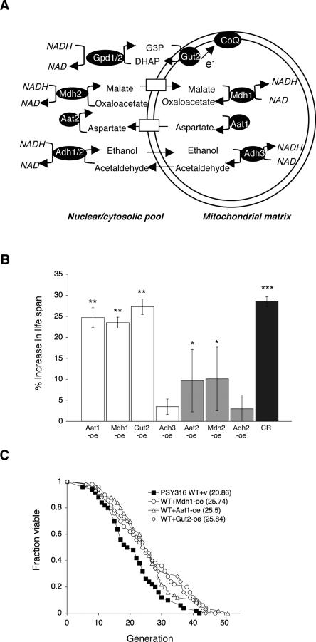
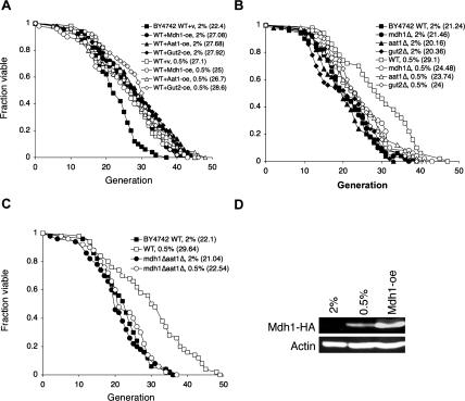
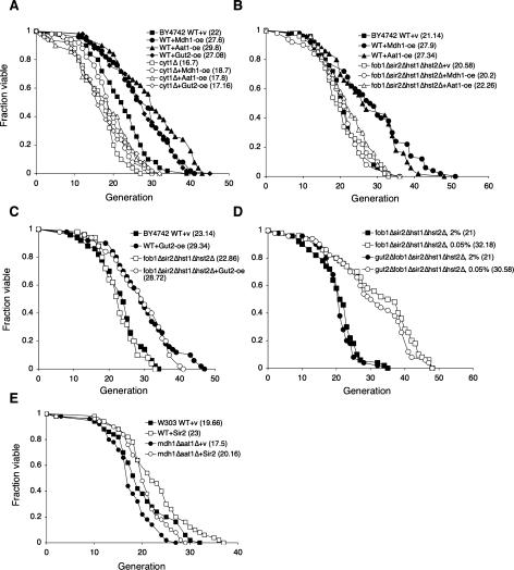
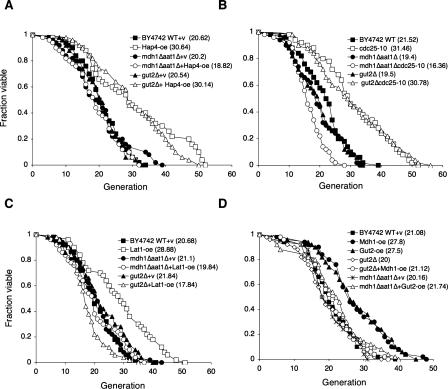
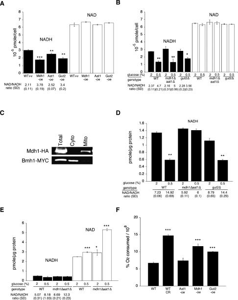
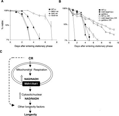
Recent studies suggest that increased mitochondrial metabolism and the concomitant decrease in NADH levels mediate calorie restriction (CR)-induced life span extension. The mitochondrial inner membrane is impermeable to NAD (nicotinamide adenine dinucleotide, oxidized form) and NADH, and it is unclear how CR relays increased mitochondrial metabolism to multiple cellular pathways that reside in spatially distinct compartments. Here we show that the mitochondrial components of the malate-aspartate NADH shuttle (Mdh1 [malate dehydrogenase] and Aat1 [aspartate amino transferase]) and the glycerol-3-phosphate shuttle (Gut2, glycerol-3-phosphate dehydrogenase) are novel longevity factors in the CR pathway in yeast. Overexpressing Mdh1, Aat1, and Gut2 extend life span and do not synergize with CR. Mdh1 and Aat1 overexpressions require both respiration and the Sir2 family to extend life span. The mdh1Deltaaat1Delta double mutation blocks CR-mediated life span extension and also prevents the characteristic decrease in the NADH levels in the cytosolic/nuclear pool, suggesting that the malate-aspartate shuttle plays a major role in the activation of the downstream targets of CR such as Sir2. Overexpression of the NADH shuttles may also extend life span by increasing the metabolic fitness of the cells. Together, these data suggest that CR may extend life span and ameliorate age-associated metabolic diseases by activating components of the NADH shuttles.


Figures

Figure 1.
Figure 1.
Overexpressing components of the NADH shuttle systems extend life span. (A) Schematic presentation of the yeast NADH shuttle systems that balance the NAD/NADH ratio between the cytosolic and the mitochondrial pools. For example, in the case of the ethanol–acetaldehyde shuttle, an increase in the mitochondrial NAD/NADH ratio leads to the production of acetaldehyde through mitochondrial alcohol dehydrogenases (Adh3). Mitochondrial acetaldehyde diffuses freely to the cytosol, which is then reduced to ethanol via cytosolic alcohol dehydrogenases (Adh1/2), resulting in an increase in the cytosolic NAD/NADH ratio. In the malate–aspartate shuttle, an increase in the mitochondrial NAD/NADH ratio leads to the production of aspartate from malate through the malate dehydrogenase (Mdh1) and the aspartate aminase (Aat1). Aspartate is transported to the cytosol via the Agc1 carrier, which is then converted to malate by cytosolic malate dehydrogenase (Mdh2) and aspartate aminase (Aat2), resulting in an increase in the cytosolic NAD/NADH ratio. Cytosolic malate enters mitochondria though specific carriers. In the glycerol-3-phosphate shuttle, the mitochondrial glycerol-3-phosphate (G-3-P) dehydrogenase (Gut2) converts G-3-P to DHAP (dihydroxyacetone phosphate), resulting in an increase in the cytosoilc NAD/NADH ratio through the action of the cytosolic G-3-P dehydrogenases (Gpd1/2). Gut2 directly feeds an electron (e) from G-3-P to coenzyme Q (CoQ), supporting electron transport chain activity. (B) Overexpressing components of the NADH shuttle systems extend life span. Life span analysis of the wild-type cells carrying a control vector and various overexpression DNA constructs. Percent increase in life span relative to wild-type control grown on 2% glucose media is shown. The results show the average of three independent experiments. Error bars denote standard deviations. P values were calculated using the Student’s t test. (*) P < 0.05; (**) P < 0.005; (***) P < 0.001. (C). Mdh1, Aat1, and Gut2 overexpressions also extend life span in a different strain background. Life span analysis of cells overexpressing Mdh1, Aat1, and Gut2 in the PSY316AR strain. One set of representative data is shown. (WT) Wild-type control; (CR) 0.5% glucose; (v) empty vector; (oe) overexpression.
Figure 2.
Figure 2.
CR and the NADH shuttle system function in the same pathway. (A) CR does not synergize with the overexpressions to extend life span. Life span analysis of cells overexpressing Mdh1, Aat1, and Gut2 grown under normal and CR (0.5% glucose) conditions. (B) Deletions of single components of the NADH shuttle systems partially inhibit CR-mediated life span extension. (C) Complete inactivation of the malate–aspartate shuttle totally abolishes CR-induced life span extension. (D) CR (0.5% glucose) is sufficient to induce expression of the shuttle component. Western blot analysis of the Mdh1 protein levels in cells grown in 2% and 0.5% glucose. Mdh1 is fused to an HA epitope tag and visualized with the anti-HA antiserum. (WT) Wild-type control; (oe) overexpression. One set of representative data is shown.
Figure 3.
Figure 3.
The requirement of respiration and the Sir2 family in the NADH shuttle overexpression induced life span extension. (A) Mdh1, Aat1, and Gut2 overexpressions require Cyt1 for life span extension. Life span analysis of the wild-type cells, the cyt1Δ mutant, and the cyt1Δ mutant overexpressing Mdh1, Aat1, and Gut2 grown in 2% glucose. (B) Mdh1 and Aat1 overexpression require the Sir2 family to extend life span. Life span analysis of the wild-type, Mdh1-oe, and Aat1-oe cells, the fob1Δsir2Δhst1Δhst2Δ quadruple mutant, and the fob1Δsir2Δhst1Δhst2Δ mutant with Mdh1-oe and Aat1-oe on 2% glucose media. (C) Gut2 overexpression does not require the Sir2 family to extend life span. Life span analysis of the wild-type and Gut2-oe cells, the fob1Δsir2Δhst1Δhst2Δ quadruple mutant, and the fob1Δsir2Δhst1Δhst2Δ mutant with Gut2-oe on 2% glucose media. (D) To extend life span, 0.05% glucose does not require Gut2. Life span analysis of the fob1Δsir2Δhst1Δhst2Δ quadruple mutant and the gut2Δfob1Δsir2Δhst1Δhst2Δ quintuple mutant cells grown in 0.05% and 2% glucose. (E) Expressing one extra copy of Sir2 requires the malate–aspartate shuttle for maximum life span extension in the W303AR strain background. (WT) Wild-type control; (v) empty vector control; (oe) overexpression. One set of representative data is shown.
Figure 4.
Figure 4.
CR genetic models and the NADH shuttle. (A) Overexpression of Hap4 requires the malate–aspartate shuttle for life span extension. Life span analysis of the wild-type and Hap4-oe cells, the mdh1Δaat1Δ double mutant, and gut2Δ single mutant with and without Hap4-oe on 2% glucose media. (B) The cdc25-10 mutation requires the malate–aspartate shuttle for life span extension. Life span analysis of the wild-type and cdc25-10 mutant cells, the mdh1Δaat1Δ double mutant, and gut2Δ single mutant with and without the cdc25-10 mutation on 2% glucose media. (C) Lat1 overexpression requires both the malate–aspartate shuttle and the glycerol-3-phosphate shuttle to extend life span. (D) Mdh1 overexpression requires the glycerol-3-phosphate shuttle to extend life span and Gut2 overexpression requires the malate–aspartate shuttle to extend life span. (WT) Wild-type control; (v) empty vector control; (oe) overexpression. One set of representative data is shown.
Figure 5.
Figure 5.
Role of the NADH shuttles in the regulation of intracellular NAD/NADH levels. Measurements of the intracellular NAD and NADH levels in wild-type cells, cells overexpressing the shuttle components, and cells carrying deletions in the shuttle components grown in 2% and 0.5% glucose. (A) Mdh1, Aat1, and Gut2 overexpression decreases the total NADH levels. (B) Deletions of the shuttle components do not abolish the decrease in the total NADH levels induced by CR. (C) Western blot analysis of total cellular, cytosolic, and mitochondrial fractions used for NAD/NADH ratio assay analysis. (D) Inactivation of the malate–aspartate shuttle abolishes the decrease in the NADH levels in the cytosolic/nuclear pools induced by CR. (E) Inactivation of the malate–aspartate shuttle further increases the NAD/NADH ratio in mitochondria under CR. (F) Gut2 and Mdh1 overexpressions significantly increase oxygen consumption rates compared with wild-type cells grown in 2% glucose. (WT) BY4742 wild type; (v) empty vector control; (oe) overexpression; (SD) standard deviations. One representative set of three independent experiments, each conducted in quadruplicate (A,B) or triplicate (DF), is shown. Error bars denote standard deviations. P values were calculated using the Student’s t test. (*) P < 0.05; (**) P < 0.005; (***) P < 0.001.
Figure 6.
Figure 6.
The NADH shuttle systems and life span regulation. (A) Mdh1 and Aat1 overexpressions extend CLS. (B) The malate–aspartate shuttle is required for maximum CLS extension induced by CR. Fractions of viable wild-type, Mdh1-oe, Aat1-oe, Gut2-oe, mdh1Δaat1Δ double mutant, and gut2Δ single mutant cells were determined from cultures grown to stationary phase. One representative set of three independent experiments, each conducted in triplicate, is shown. Error bars denote standard deviations. (WT) BY4742 wild type; (CR) 0.5% glucose; (v) empty vector control; (oe) overexpression. (C) A model for the role of the NADH shuttle in CR. Multiple CR pathways act to affect longevity in yeast. CR activates mitochondrial respiration, which is required for 0.5% CR and CR mimic-induced life span extension. A functional malate–aspartate shuttle is also required for CR-induced life span extension. We propose that under CR, the malate–aspartate shuttle transmits the increase in the NAD/NADH ratio from the mitochondrial pool to the cytosolic/nuclear pool. The increased NAD/NADH ratio in the cytosolic/nuclear pool then activates other longevity factors such as the Sir2 family to extend life span. Overexpression of the shuttle components (such as Gut2) may also extend life span by maintaining mitochondrial activity, thereby increasing metabolic fitness of the cell.

Similar articles

Cited by

References

    1. Aguilaniu H., Gustafsson L., Rigoulet M., Nystrom T. Asymmetric inheritance of oxidatively damaged proteins during cytokinesis. Science. 2003;299:1751–1753. - PubMed
    1. Albertyn J., Hohmann S., Thevelein J.M., Prior B.A. GPD1, which encodes glycerol-3-phosphate dehydrogenase, is essential for growth under osmotic stress in Saccharomyces cerevisiae, and its expression is regulated by the high-osmolarity glycerol response pathway. Mol. Cell. Biol. 1994;14:4135–4144. - PMC - PubMed
    1. Anderson R.M., Bitterman K.J., Wood J.G., Medvedik O., Sinclair D.A. Nicotinamide and PNC1 govern lifespan extension by calorie restriction in Saccharomyces cerevisiae. Nature. 2003;423:181–185. - PMC - PubMed
    1. Bakker B.M., Bro C., Kotter P., Luttik M.A., van Dijken J.P., Pronk J.T. The mitochondrial alcohol dehydrogenase Adh3p is involved in a redox shuttle in Saccharomyces cerevisiae. J. Bacteriol. 2000;182:4730–4737. - PMC - PubMed
    1. Bakker B.M., Overkamp K.M., van Maris A.J., Kotter P., Luttik M.A., van Dijken J.P., Pronk J.T. Stoichiometry and compartmentation of NADH metabolism in Saccharomyces cerevisiae. FEMS Microbiol. Rev. 2001;25:15–37. - PubMed

Publication types

MeSH terms