. 2008 Apr 1;22(7):945-53.
doi: 10.1101/gad.1645008.

Control of cell fate by the formation of an architecturally complex bacterial community

Affiliations

Control of cell fate by the formation of an architecturally complex bacterial community

Hera Vlamakis et al. Genes Dev. .

Abstract

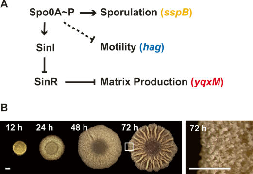
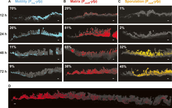
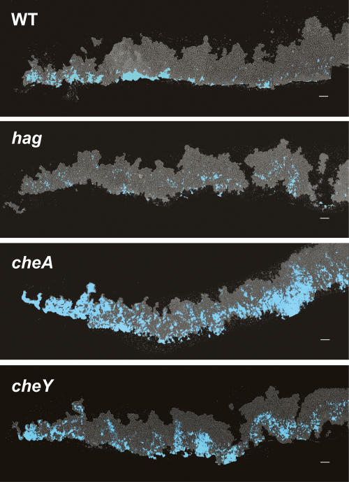
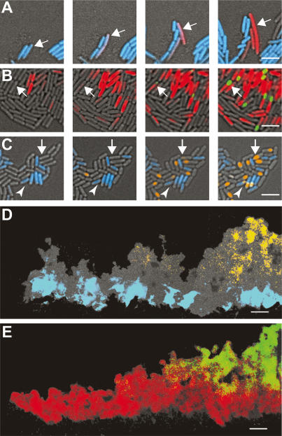
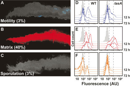
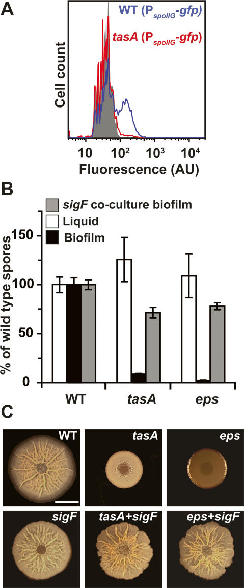
Bacteria form architecturally complex communities known as biofilms in which cells are held together by an extracellular matrix. Biofilms harbor multiple cell types, and it has been proposed that within biofilms individual cells follow different developmental pathways, resulting in heterogeneous populations. Here we demonstrate cellular differentiation within biofilms of the spore-forming bacterium Bacillus subtilis, and present evidence that formation of the biofilm governs differentiation. We show that motile, matrix-producing, and sporulating cells localize to distinct regions within the biofilm, and that the localization and percentage of each cell type is dynamic throughout development of the community. Importantly, mutants that do not produce extracellular matrix form unstructured biofilms that are deficient in sporulation. We propose that sporulation is a culminating feature of biofilm formation, and that spore formation is coupled to the formation of an architecturally complex community of cells.


Figures

Figure 1.
Figure 1.
(A) Working model of the regulation of differentiation. Intracellular changes in the concentration of Spo0A∼P regulate differential gene expression. While sporulation requires high Spo0A∼P levels, the expression of SinI is achieved at lower levels of Spo0A∼P. When expressed, SinI antagonizes SinR, which directly represses genes involved in extracellular matrix production. Spo0A∼P is also involved in repressing motility. Solid lines indicate known regulatory mechanisms while the dashed line indicates an unknown mechanism. The sspB, hag, and yqxM genes are expressed in sporulating, motile, and matrix-producing cells, respectively. (B) Top view of biofilm development over time. The right panel is magnified to highlight the aerial structures observed at 72 h. The boxed region indicates the portion of the biofilm displayed later in Figure 3. Bars, 1 mm.
Figure 2.
Figure 2.
Flow cytometry analysis of cells expressing Phag-yfp reporters (motility, blue shading) (A), PyqxM-yfp reporters (matrix-production, red shading) (B), or PsspB-yfp reporters (sporulation, orange shading) (C). Expression was followed over a period of 72 h. In gray is the peak for control cells with no yfp reporter. The Y-axis represents cell counts for each strain (30,000 cells were counted) at 12-, 24-, 48-, and 72-h time points. The X-axis is arbitrary units (AU) of fluorescence in a logarithmic scale.
Figure 3.
Figure 3.
Spatiotemporal analysis of differentiation within a biofilm. Vertical thin sections of biofilms harboring the indicated reporter fusions. (Left) Biofilms were frozen at the indicated times of development prior to cryosectioning and fixation. Images represent only partial colonies approximately encompassing the region magnified in Figure 1B. The edge of the colony is shown at the left of the image, the agar surface is at the bottom. Transmitted light images (cells appear gray on the black background) were overlaid with fluorescence images that were false-colored blue for motility (A), red for matrix production (B), and orange for sporulation (C). The percentage of fluorescent cells was obtained by flow cytometry, and is indicated in the top left of each image. Percentages represent the average obtained from three independent experiments. (D) Image of 72-h thin section from B encompassing the edge to the center of the colony. Bars, 50 μm.
Figure 4.
Figure 4.
Motility is required for proper localization of Phag-cfpexpressing cells (blue). Vertical thin sections of biofilms harvested at 48 h of development. hag encodes flagellin, and cheA and cheY are necessary for chemotaxis. Colony orientation is as in Figure 3. Bar, 50 μm.
Figure 5.
Figure 5.
Developmental history of cells within a biofilm. Time-lapse images of cells harboring dual reporter fusions. (A) Images of cells harboring Phag-cfp (motile cells, blue) and PyqxM-yfp (matrix-producing cells, red) were taken every 1 h. Arrow indicates a motile cell transitioning to matrix-producing cell. (B) Images of cells harboring PyqxM-cfp (matrix-producing cells, red) and PsspB-yfp (sporulating cells, green) were taken every 2 h. Arrow highlights a cell initiating matrix production and then transitioning to a sporulating cell. (C) Images of cells harboring Phag-cfp (motile cells, blue) and PsspB-yfp (sporulating cells, orange) were taken every 2 h. The majority of the sporulating cells arise from nonmotile cells, indicated by the white arrow. The arrowhead highlights an example of the minority of motile cells that initiate sporulation. (D,E) Thin sections of 48-h colonies from cells harboring dual reporters. Images formatted as in Figure 3. In D, motile cells (blue) appear in distinct regions relative to sporulating cells (orange), whereas in E, matrix-producing cells (red) overlap with the region of sporulating cells (green). Bars: A–C, 5 μm; D,E, 50 μm.
Figure 6.
Figure 6.
The effect of an altered extracellular matrix (tasA) on spatiotemporal regulation of gene expression. (A–C) Thin sections of 72-h tasA mutant biofilms harboring the indicated reporters. False coloring for each reporter and colony orientation is as in Figure 3. The percent of cells expressing each reporter obtained by flow cytometry is indicated in the bottom left corner of each image. Bar, 50 μm. (D–F) Dynamics of gene expression are altered in a matrix-deficient mutant. Flow cytometry analysis as in Figure 2. Wild-type (left panels) versus tasA mutant (right panels) cells obtained from biofilms harvested at 12, 24, 48, or 72 h. Gray peak indicates fluorescence intensity of cells with no fluorescent reporter. Reporters used are Phag-yfp for motility (D), PyqxM-yfp for matrix production (E), and PsspB-yfp for sporulation (F). Dashed gray lines bracket low fluorescence levels. To the left are cells not expressing the reporters, and to the right are cells expressing high levels of fluorescence.
Figure 7.
Figure 7.
The effect of an altered extracellular matrix on gene expression, sporulation, and biofilm architecture. (A) Flow cytometry analysis of PspoIIG-gfp expression as in Figure 2. Wild-type (blue line) and tasA mutant (red line) cells obtained from biofilms harvested at 36 h. Gray peak indicates fluorescence intensity of cells with no fluorescent reporter. (B) Viable spore counts comparing mutants in matrix production (tasA and eps) to wild-type percent of spores in biofilms (black bars) or liquid cultures (white bars). Sporulation was restored by coculturing the different strains with a nonsporulating (sigF) mutant (gray bars). Error bars indicate SEM. (C) Coculturing restores biofilm architecture to matrix-deficient mutants. Top view of biofilms from wild-type, tasA, and eps (top panels) and sigF and cocultures of tasA or eps cells mixed at a 1:1 ratio with a sigF mutant (bottom panels) after 72 h of incubation at 30°C. Bar, 1 cm.

References

    1. An D., Parsek M.R. The promise and peril of transcriptional profiling in biofilm communities. Curr. Opin. Microbiol. 2007;10:292–296. - PubMed
    1. Branda S.S., Gonzalez-Pastor J.E., Ben-Yehuda S., Losick R., Kolter R. Fruiting body formation by Bacillus subtilis. Proc. Natl. Acad. Sci. 2001;98:11621–11626. - PMC - PubMed
    1. Branda S.S., Chu F., Kearns D.B., Losick R., Kolter R. A major protein component of the Bacillus subtilis biofilm matrix. Mol. Microbiol. 2006;59:1229–1238. - PubMed
    1. Burbulys D., Trach K.A., Hoch J.A. Initiation of sporulation in B. subtilis is controlled by a multicomponent phosphorelay. Cell. 1991;64:545–552. - PubMed
    1. Chai Y., Chu F., Kolter R., Losick R. Bistability and biofilm formation in Bacillus subtilis. Mol. Microbiol. 2007;67:254–263. - PMC - PubMed

Publication types