

The anatomy of first-episode and chronic schizophrenia: an anatomical likelihood estimation meta-analysis
- PMID: 18381902
- PMCID: PMC2873788
- DOI: 10.1176/appi.ajp.2008.07101562
The anatomy of first-episode and chronic schizophrenia: an anatomical likelihood estimation meta-analysis
Abstract
Objective: The authors sought to map gray matter changes in first-episode schizophrenia and to compare these with the changes in chronic schizophrenia. They postulated that the data would show a progression of changes from hippocampal deficits in first-episode schizophrenia to include volume reductions in the amygdala and cortical gray matter in chronic schizophrenia.
Method: A systematic search was conducted for voxel-based structural MRI studies of patients with first-episode schizophrenia and chronic schizophrenia in relation to comparison groups. Meta-analyses of the coordinates of gray matter differences were carried out using anatomical likelihood estimation. Maps of gray matter changes were constructed, and subtraction meta-analysis was used to compare them.
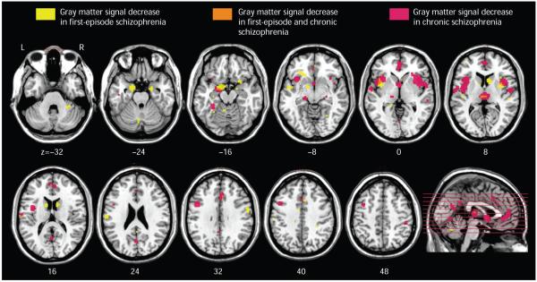
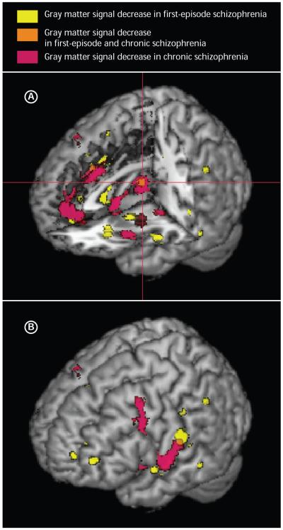
Results: A total of 27 articles were identified for inclusion in the meta-analyses. A marked correspondence was observed in regions affected by both first-episode schizophrenia and chronic schizophrenia, including gray matter decreases in the thalamus, the left uncus/amygdala region, the insula bilaterally, and the anterior cingulate. In the comparison of first-episode schizophrenia and chronic schizophrenia, decreases in gray matter volume were detected in first-episode schizophrenia but not in chronic schizophrenia in the caudate head bilaterally; decreases were more widespread in cortical regions in chronic schizophrenia.
Conclusions: Anatomical changes in first-episode schizophrenia broadly coincide with a basal ganglia-thalamocortical circuit. These changes include bilateral reductions in caudate head gray matter, which are absent in chronic schizophrenia. Comparing first-episode schizophrenia and chronic schizophrenia, the authors did not find evidence for the temporolimbic progression of pathology from hippocampus to amygdala, but there was evidence for progression of cortical changes.
Figures
Comment in
-
Neural networks in schizophrenia.Am J Psychiatry. 2008 Aug;165(8):937-9. doi: 10.1176/appi.ajp.2008.08050700. Am J Psychiatry. 2008. PMID: 18676594 No abstract available.
References
-
- Steen RG, Mull C, McClure R, Hamer RM, Lieberman JA. Brain volume in first-episode schizophrenia: systematic review and meta-analysis of magnetic resonance imaging studies. Br J Psychiatry. 2006;188:510–518. - PubMed
-
- Vita A, De Peri L, Silenzi C, Dieci M. Brain morphology in first-episode schizophrenia: a meta-analysis of quantitative magnetic resonance imaging studies. Schizophr Res. 2006;82:75–88. - PubMed
-
- Vita A, de Peri L. Hippocampal and amygdala volume reductions in first-episode schizophrenia. Br J Psychiatry. 2007;190:271. - PubMed
-
- Lawrie SM, Abukmeil SS. Brain abnormality in schizophrenia: a systematic and quantitative review of volumetric magnetic resonance imaging studies. Br J Psychiatry. 1998;172:110–120. - PubMed
-
- Wright IC, Rabe-Hesketh S, Woodruff PWR, David AS, Murray RM, Bullmore ET. Meta-analysis of regional brain volumes in schizophrenia. Am J Psychiatry. 2000;157:16–25. - PubMed
Publication types
MeSH terms
Grants and funding
LinkOut - more resources
Full Text Sources
Other Literature Sources
Medical
