Comparative Study
. 2008 Apr 1;14(7):1938-46.
doi: 10.1158/1078-0432.CCR-07-4082.

Heterogeneity of breast cancer metastases: comparison of therapeutic target expression and promoter methylation between primary tumors and their multifocal metastases

Affiliations
Comparative Study

Heterogeneity of breast cancer metastases: comparison of therapeutic target expression and promoter methylation between primary tumors and their multifocal metastases

Julie M Wu et al. Clin Cancer Res. .

Abstract

Purpose: A comprehensive comparison of biomarker expression between patients' primary breast carcinoma (PBC) and their metastatic breast carcinomas (MBC) has not been done.

Experimental design: We did rapid autopsies (postmortem intervals, 1-4 hours) on 10 consenting patients who died of MBC. We constructed single-patient tissue microarrays from the patients' archived PBC and multiple different MBCs harvested at autopsy, which were immunohistochemically labeled for multiple biomarkers. Methylation of multiple gene promoters was assessed quantitatively on dissected PBC and MBC samples.

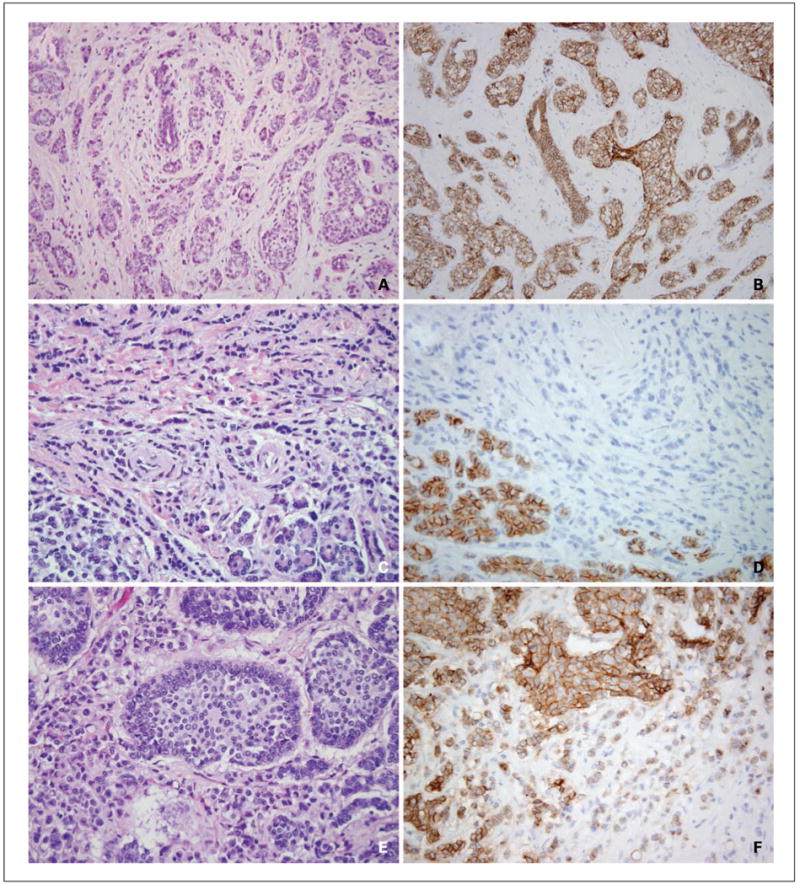
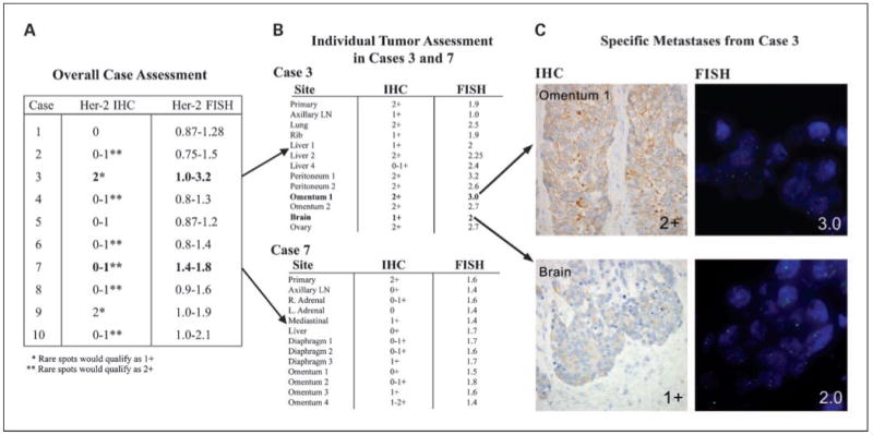
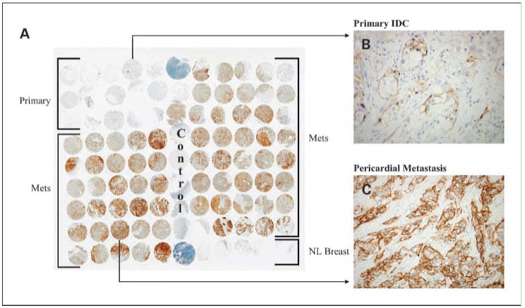
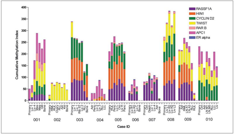
Results: Extensive heterogeneity was observed between PBC and their paired MBC, as well as among multiple MBC from the same patient. Estrogen and progesterone receptors tended to be uniformly down-regulated in metastases. E-cadherin was down-regulated in a subset of the MBC of one case. Variable overexpression in MBC compared with the PBC was observed for cyclooxygenase-2 (five cases), epidermal growth factor receptor (EGFR; four cases), MET (four cases), and mesothelin (four cases). No case strongly overexpressed HER-2/neu by immunohistochemistry, but eight cases showed variable protein expression ranging from negative to equivocal (2+) in different MBC. In one case, variable low-level HER-2/neu gene amplification was found. EGFR and MET overexpression were restricted to the four basal-type cancers. EGFR protein overexpression did not correlate with EGFR gene amplification. Multigene promoter hypermethylation of RASSF1a, HIN1, cyclin D2, Twist, estrogen receptor alpha, APC1, and RARbeta was overall very similar in the PBC and all MBCs in all cases.

Conclusions: Therapeutic targets identified in the PBC or even some MBC may not reflect targets present in all metastatic sites.


Figures

Fig. 1
Fig. 1
Variable morphology and E-cadherin immunohistochemistry of MBC5 metastases. The primary tumor (A) showed ductal morphology in that it grew as cords and solid nests and accordingly showed intact E-cadherin protein expression by immunohistochemistry (B). The majority of metastases, including pancreatic metastases (C), showed lobular morphology and accordingly showed loss of E-cadherin protein expression (D). Note that the native pancreatic epithelium (bottom left) labels for E-cadherin, providing an internal control. A pericardial metastasis (E) showed predominant nested morphology consistent with ductal differentiation, and E-cadherin was largely intact in this metastasis (F). Note the additional cord-like area of signet ring cells (E, bottom left) which showed diminished E-cadherin expression (F, bottom right).
Fig. 2
Fig. 2
HER-2/neu analysis of primaries and metastases. A, range of HER-2/neu immunohistochemistry labeling scores for each case, along with range of HER-2/neu gene amplification ratios. B, range of HER-2/neu immunohistochemistry labeling scores for each metastases of cases 3 and 7 compared with the primary, along with corresponding HER-2/neu gene amplification ratios. Note the variable low-level amplification in MBC3, whereas the focal 2+ labeling by immunohistochemistry in MBC7 does not correlate with amplification. C, HER-2/neu assessment of selected individual tumor spots from MBC3 omental metastases 1 shows 2+ staining by immunohistochemistry and is amplified (ratio, 3). Brain metastases shows 1+ labeling by immunohistochemistry but is borderline amplified (ratio, 2).
Fig. 3
Fig. 3
Striking EGFR protein up-regulation in metastases of MBC10. A, low power view of TMA labeled for EGFR protein by immunohistochemistry. Note the strong labeling of all metastases (rows 4-9, spots 1-5 and rows 1-8, spots 7-11) compared with normal breast (row 9, spots 7-11) and the primary infiltrating ductal carcinoma (rows 1-3, spots 1-5). B, individual spot of primary tumor, showing focal labeling for EGFR. C, individual spot of pericardial metastasis, showing diffuse labeling for EGFR.
Fig. 4
Fig. 4
Cumulative promoter hypermethylation of RASSF1A, HIN1, CYCLIN D2, TWIST, RARβ, APC1, and ERα in lesions of primary and metastatic breast cancer. Scores for cumulative methylation were obtained by quantitative multiplex methylation-specific PCR assay in liver (Liv), mesentery (Mes), gall bladder (GB), bone, lung (RUL, lobes of right upper; RML, right middle; RLL, right lower; LLL, left lower), ovary, adrenal (Adr), kidney (Kid), rib soft tissue (ST), pericardium (Peric), omentum (Om), diaphragm (Diaph), and lymph node [LN; abdominal (A) and mediastinal (M)]. Quantitive multiplex methylation-specific PCR analysis of both unmethylated and methylated gene promoters was done. The percentage of methylation of each gene was calculated as 100 × [no. of copies methylated DNA/(no. of copies unmethylated + methylated DNA)]. The cumulative methylation index score was obtained as the sum of the percentage of methylation for each of the seven genes. Results are plotted as stacked bar graphs where the column height reflects the total cumulative methylation; for a maximum possible of 700 cumulative units (100% × 7 genes), the colored segment corresponding to the percentage of methylation of each gene color-coded in the top right legend. Ten patients (MBC1-MBC10) were evaluated. Data is also shown in Supplementary Table S5.

Comment in

References

    1. Paget S. The distribution of secondary growths in cancer of the breast. Cancer Metastasis Rev. 1989;8:98–101. - PubMed
    1. Weigelt B, Glas AM, Wessels LF, Witteveen AT, Peterse JL, van't Veer LJ. Gene expression profiles of primary breast tumors maintained in distant metastases. Proc Natl Acad Sci U S A. 2003;100:15901–5. - PMC - PubMed
    1. Kang Y, Siegel PM, Shu W, et al. A multigenic program mediating breast cancer metastasis to bone. Cancer Cell. 2003;3:537–49. - PubMed
    1. Minn AJ, Gupta GP, Siegel PM, et al. Genes that mediate breast cancer metastasis to lung. Nature. 2005;436:518–24. - PMC - PubMed
    1. Al-Hajj M, Wicha MS, Benito-Hernandez A, Morrison SJ, Clarke MF. Prospective identification of tumorigenic breast cancer cells. Proc Natl Acad Sci U S A. 2003;100:3983–8. - PMC - PubMed

Publication types

Substances