

A Role for PPARbeta/delta in Ocular Angiogenesis
- PMID: 18382612
- PMCID: PMC2276600
- DOI: 10.1155/2008/825970
A Role for PPARbeta/delta in Ocular Angiogenesis
Abstract
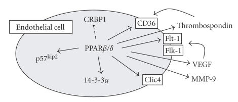
The uses of highly selective PPARbeta/delta ligands and PPARbeta/delta knockout mice have shown a direct ability of PPARbeta/delta to regulate angiogenesis in vitro and in vivo in animal models. PPARbeta/delta ligands induce the proangiogenic growth factor VEGF in many cells and tissues, though its actions in the eye are not known. However, virtually, all tissue components of the eye express PPARbeta/delta. Both angiogenesis and in particular VEGF are not only critical for the development of the retina, but they are also a central component in many common pathologies of the eye, including diabetic retinopathy and age-related macular degeneration, the most common causes of blindness in the Western world. This review, therefore, will discuss the recent evidence of PPARbeta/delta-mediated angiogenesis and VEGF release in the context of ocular disorders.
Figures

References
-
- Moraes LA, Piqueras L, Bishop-Bailey D. Peroxisome proliferator-activated receptors and inflammation. Pharmacology & Therapeutics. 2006;110(3):371–385. - PubMed
-
- Dreyer C, Krey G, Keller H, Givel F, Helftenbein G, Wahli W. Control of the peroxisomal β-oxidation pathway by a novel family of nuclear hormone receptors. Cell. 1992;68(5):879–887. - PubMed
-
- Mukherjee R, Jow L, Croston GE, Paterniti JR., Jr Identification, characterization, and tissue distribution of human peroxisome proliferator-activated receptor (PPAR) isoforms PPARγ2 versus PPARγ1 and activation with retinoid X receptor agonists and antagonists. Journal of Biological Chemistry. 1997;272(12):8071–8076. - PubMed
-
- Vosper H, Patel L, Graham TL, et al. The peroxisome proliferator-activated receptor δ promotes lipid accumulation in human macrophages. Journal of Biological Chemistry. 2001;276(47):44258–44265. - PubMed
LinkOut - more resources
Full Text Sources
Other Literature Sources
