

Prevention of Oxidative Stress-Induced Retinal Pigment Epithelial Cell Death by the PPARgamma Agonists, 15-Deoxy-Delta 12, 14-Prostaglandin J(2)
- PMID: 18382621
- PMCID: PMC2276681
- DOI: 10.1155/2008/720163
Prevention of Oxidative Stress-Induced Retinal Pigment Epithelial Cell Death by the PPARgamma Agonists, 15-Deoxy-Delta 12, 14-Prostaglandin J(2)
Abstract
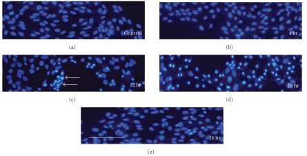
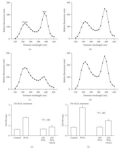
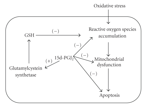
Cellular oxidative stress plays an important role in retinal pigment epithelial (RPE) cell death during aging and the development of age-related macular degeneration. Early reports indicate that during phagocytosis of rod outer segments, there is an increase of RPE oxidative stress and an upregulation of PPARgamma mRNA in these cells. These studies suggest that activation of PPARgamma may modulate cellular oxidative stress. This paper presents a brief review of recent studies that investigate RPE oxidative stress under various experimental conditions. This is followed by a detailed review on those reports that examine the protective effect of the natural PPARgamma ligand, 15d-PGJ(2), against RPE oxidative stress. This agent can upregulate glutathione and prevent oxidant-induced intracellular reactive oxygen species accumulation, mitochondrial depolarization, and apoptosis. The cytoprotective effect of this agent, however, is not shared by other PPARgamma agonists. Nonetheless, this property of 15d-PGJ(2) may be useful in future development of pharmacological tools against retinal diseases caused by oxidative stress.
Figures
Similar articles
-
15-Deoxy-Δ(12,14)-prostaglandin J(2) attenuates the biological activities of monocyte/macrophage cell lines.Eur J Cell Biol. 2012 Aug;91(8):654-61. doi: 10.1016/j.ejcb.2012.03.004. Epub 2012 May 4. Eur J Cell Biol. 2012. PMID: 22560326
-
The 15-deoxy-δ12,14-prostaglandin J2 inhibits LPS‑stimulated inflammation via enhancement of the platelet‑activating factor acetylhydrolase activity in human retinal pigment epithelial cells.Int J Mol Med. 2014 Feb;33(2):449-56. doi: 10.3892/ijmm.2013.1588. Epub 2013 Dec 13. Int J Mol Med. 2014. PMID: 24337644
-
15-Deoxy-Delta12,14-prostaglandin J(2) induces death receptor 5 expression through mRNA stabilization independently of PPARgamma and potentiates TRAIL-induced apoptosis.Mol Cancer Ther. 2006 Jul;5(7):1827-35. doi: 10.1158/1535-7163.MCT-06-0023. Mol Cancer Ther. 2006. PMID: 16891469
-
Peroxisome proliferator-activated receptor gamma (PPARgamma) ligands as bifunctional regulators of cell proliferation.Biochem Pharmacol. 2003 Oct 15;66(8):1381-91. doi: 10.1016/s0006-2952(03)00488-x. Biochem Pharmacol. 2003. PMID: 14555212 Review.
-
The impact of oxidative stress and inflammation on RPE degeneration in non-neovascular AMD.Prog Retin Eye Res. 2017 Sep;60:201-218. doi: 10.1016/j.preteyeres.2017.03.002. Epub 2017 Mar 20. Prog Retin Eye Res. 2017. PMID: 28336424 Free PMC article. Review.
Cited by
-
Expression of peroxisome proliferator-activated receptor γ in rat retina during development.Int J Ophthalmol. 2015 Feb 18;8(1):52-6. doi: 10.3980/j.issn.2222-3959.2015.01.09. eCollection 2015. Int J Ophthalmol. 2015. PMID: 25709907 Free PMC article.
-
Modulation of diabetic retinopathy pathophysiology by natural medicines through PPAR-γ-related pharmacology.Br J Pharmacol. 2012 Jan;165(1):4-19. doi: 10.1111/j.1476-5381.2011.01411.x. Br J Pharmacol. 2012. PMID: 21480863 Free PMC article. Review.
-
Emerging roles for nuclear receptors in the pathogenesis of age-related macular degeneration.Cell Mol Life Sci. 2014 Dec;71(23):4617-36. doi: 10.1007/s00018-014-1709-x. Epub 2014 Aug 26. Cell Mol Life Sci. 2014. PMID: 25156067 Free PMC article. Review.
-
Triamcinolone acetonide prevents oxidative stress-induced tight junction disruption of retinal pigment epithelial cells.Graefes Arch Clin Exp Ophthalmol. 2009 May;247(5):641-9. doi: 10.1007/s00417-009-1041-6. Epub 2009 Feb 3. Graefes Arch Clin Exp Ophthalmol. 2009. PMID: 19189116
-
Leveraging Nuclear Receptors as Targets for Pathological Ocular Vascular Diseases.Int J Mol Sci. 2020 Apr 21;21(8):2889. doi: 10.3390/ijms21082889. Int J Mol Sci. 2020. PMID: 32326149 Free PMC article. Review.
References
-
- O'Connell SR, Bressler NM. Age-related macular degeneration. In: Regillo CD, Brown GC, Flynn HW Jr., editors. Vitreoretinal Disease: The Essentials. New York, NY, USA: Thieme Medical Publishers; 1999. pp. 213–240.
-
- Cai J, Nelson KC, Wu M, Sternberg P, Jr., Jones DP. Oxidative damage and protection of the RPE. Progress in Retinal and Eye Research. 2000;19(2):205–221. - PubMed
-
- Del Priore LV, Kuo Y-H, Tezel TH. Age-related changes in human RPE cell density and apoptosis proportion in situ. Investigative Ophthalmology & Visual Science. 2002;43(10):3312–3318. - PubMed
-
- Dunaief JL, Dentchev T, Ying G-S, Milam AH. The role of apoptosis in age-related macular degeneration. Archives of Ophthalmology. 2002;120(11):1435–1442. - PubMed
LinkOut - more resources
Full Text Sources
