. 2008 Apr 2;3(4):e1846.
doi: 10.1371/journal.pone.0001846.

Cell death by SecTRAPs: thioredoxin reductase as a prooxidant killer of cells

Affiliations

Cell death by SecTRAPs: thioredoxin reductase as a prooxidant killer of cells

Karin Anestål et al. PLoS One. .

Abstract

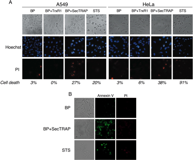
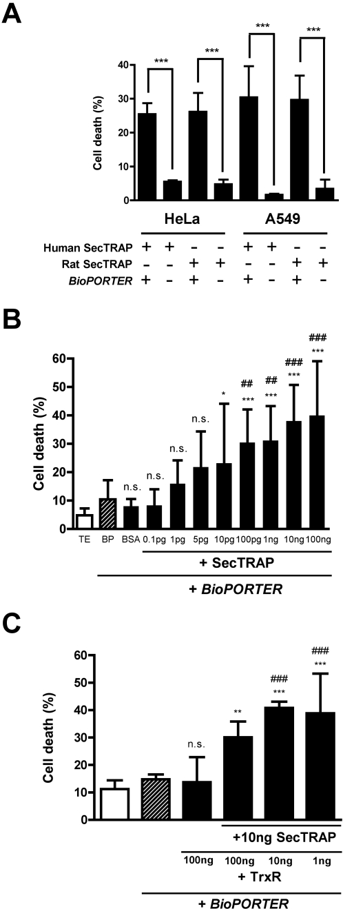
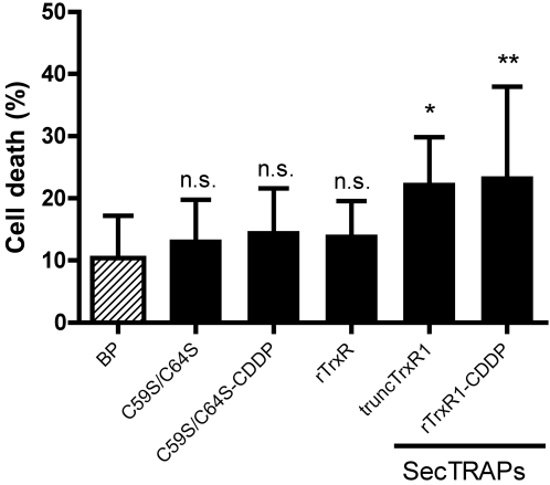
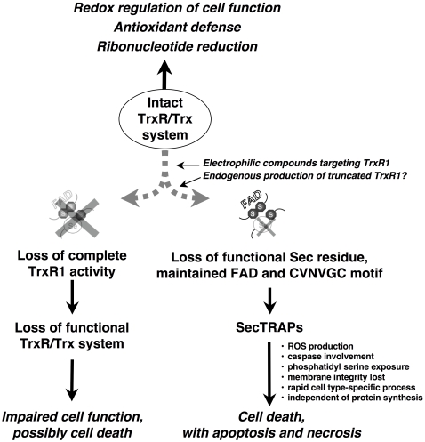
Background: SecTRAPs (selenium compromised thioredoxin reductase-derived apoptotic proteins) can be formed from the selenoprotein thioredoxin reductase (TrxR) by targeting of its selenocysteine (Sec) residue with electrophiles, or by its removal through C-terminal truncation. SecTRAPs are devoid of thioredoxin reductase activity but can induce rapid cell death in cultured cancer cell lines by a gain of function.

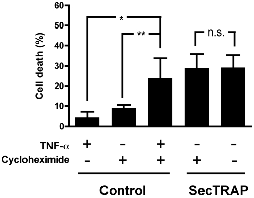
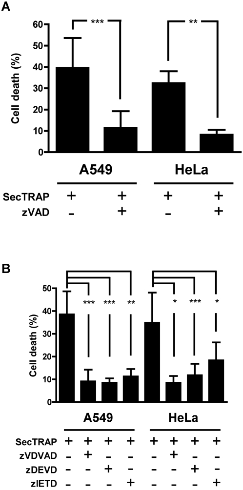
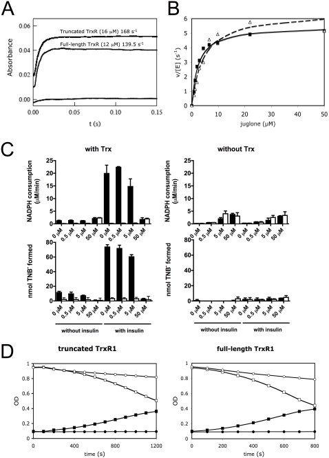
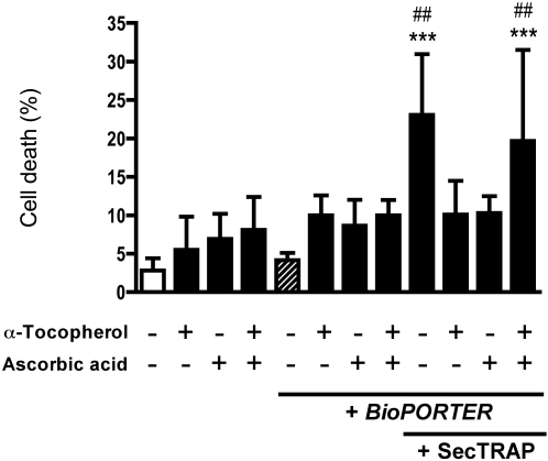
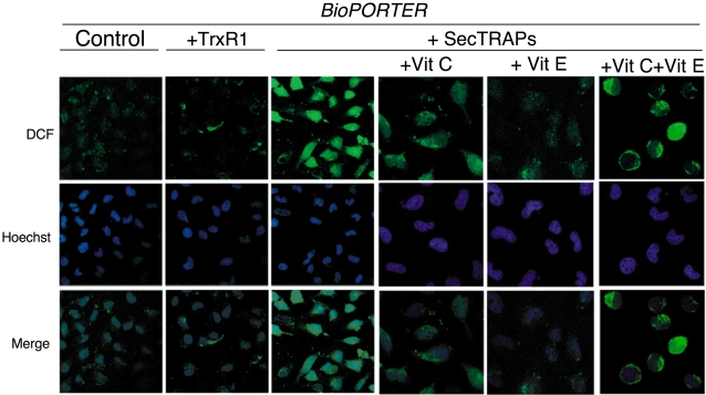
Principal findings: Both human and rat SecTRAPs killed human A549 and HeLa cells. The cell death displayed both apoptotic and necrotic features. It did not require novel protein synthesis nor did it show extensive nuclear fragmentation, but it was attenuated by use of caspase inhibitors. The redox active disulfide/dithiol motif in the N-terminal domain of TrxR had to be maintained for manifestation of SecTRAP cytotoxicity. Stopped-flow kinetics showed that NADPH can reduce the FAD moiety in SecTRAPs at similar rates as in native TrxR and purified SecTRAPs could maintain NADPH oxidase activity, which was accelerated by low molecular weight substrates such as juglone. In a cellular context, SecTRAPs triggered extensive formation of reactive oxygen species (ROS) and consequently antioxidants could protect against the cell killing by SecTRAPs.

Conclusions: We conclude that formation of SecTRAPs could contribute to the cytotoxicity seen upon exposure of cells to electrophilic agents targeting TrxR. SecTRAPs are prooxidant killers of cells, triggering mechanisms beyond those of a mere loss of thioredoxin reductase activity.


Conflict of interest statement

Competing Interests: The authors have declared that no competing interests exist.

Figures

Figure 1
Figure 1. SecTRAPs induce cell death and phosphatidyl serine exposure in human cancer cells.
(A) Morphological features and staining of HeLa and A549 cells after incubation for 4 h with BioPORTER (BP) alone, 100 ng full-length TrxR1/BioPORTER-complex, 100 ng SecTRAP/BioPORTER-complex or 1 µM staurosporine (STS), as indicated. Hoechst 33342 was used to visualize the shape or condensation of the nuclei and PI was used as a probe for lack of membrane integrity. Assessment of cell death was performed as described in the text. The percentage of cells denoted as dead, counting a total of 700–1000 cells in this particular experiment, is also given in italics in the lower part of the figure. (B) Exposure of phospatidylserine was evaluated after 3 h treatment of HeLa cells with either only BioPORTER, SecTRAP/BioPORTER-complex or 1 µM staurosporine, staining cells with Annexin-V and PI as described in the text. Magnification was x 40 in all panels.
Figure 2
Figure 2. Concentration dependent cell death induction by SecTRAPs in two human cancer cell lines and the effects of excess TrxR1.
(A) HeLa and A549 cells were treated with 100 ng rat or human SecTRAPs in the presence or absence of BioPORTER delivery reagent, as indicated in the figure and described in the text. A significant increase in cell death was seen in all cases where SecTRAPs were incubated with the cells in the presence of BioPORTER as compared to addition of SecTRAPs alone (***, p<0.001) (B) A SecTRAP preparation (truncated rat TrxR1) at an amount of 0.1 pg-100 ng was used for delivery into 10,000 A549 cells using BioPORTER, as described in the text. The graph shows the determined cell death (mean value±S.D.) triggered by each SecTRAP amount and significant differences to control treatments are indicated, using as controls either incubation with only TE buffer (white bar: n.s., p>0.05; *, p<0.05; **, p<0.01; ***, p<0.001) or with BioPORTER alone (dashed bar: n.s., p>0.05; ##, p<0.01; ###, p<0.001). No significant difference in cell death was seen comparing the two control treatments with each other. (C) A549 cells were treated with 100 ng full-length TrxR1 or a mixture of different amounts of TrxR1 with 10 ng SecTRAP using BioPORTER, as indicated in the figure. Differences in cell death were compared to control cells either treated with TE buffer (white bar; **, p<0.01; ***, p<0.001) or with only BioPORTER (dashed bar; ###, p<0.001). No statistically significant difference in cell death was seen between the two control treatments or in comparisons of either control with the treatment using 100 ng TrxR1 (n.s., p>0.05). In all experiments (A–C) cells were incubated for 4 h with the separate treatments and were subsequently stained with Hoechst 33342 and PI for evaluation of dead cells as described in the text.
Figure 3
Figure 3. Cell death by SecTRAPs is not dependent upon induction of protein synthesis.
HeLa cells were preincubated 12 h with TNF-α, cycloheximide or a combination of TNF-α and cycloheximide, whereupon SecTRAP/BioPORTER-complex was added as indicated and the cells were then incubated for additional 4 h. Cell death was subsequently evaluated by staining with Hoechst and PI as described. As a control experiment, an expected increase of cell death was seen when cells were treated with the combination of TNF-α and cycloheximide compared to treatment of either of these compounds alone, showing that the cycloheximide treatment had inhibited protein synthesis (see text). In contrast, cycloheximide had no effect on the cell death provoked by the SecTRAP/BioPORTER treatment, as indicated in the figure (n.s., p>0.05; *, p<0.05; **, p<0.01).
Figure 4
Figure 4. Cell death induction by SecTRAPs is prevented by caspase-2 and caspase-3/7 inhibitors.
(A) shows that the cell death provoked by SecTRAPs is significantly decreased upon preincubation of either A549 or HeLa cells for 30 min with 100 µM of the general caspase inhibitor zVAD before the SecTRAP treatment (**, p<0.01; ***, p<0.001). In (B) HeLa or A549 cells were incubated 30 min with 25 µM of inhibitors for caspase-2 (zVDVAD-fmk), caspase-3 (zDEVD-fmk) or caspase-8 (zIETD-fmk) before SecTRAP treatment. In all of these cases a significantly lower cell death was observed, as indicated in the figure, suggesting that the three caspases may be involved in propagating the cell death triggered by SecTRAPs, as further discussed in the text (*, p<0.05; **, p<0.01; ***, p<0.001).
Figure 5
Figure 5. Both SecTRAPs and TrxR1 are efficient in reducing juglone and thereby produce superoxide.
In (A) the formation of FAD-reduced disulfide charge transfer complex by NADPH (80 µM) in a SecTRAP preparation (truncated TrxR1, 16 µM subunit) and full-length TrxR1 (12 µM subunit) was analyzed with stopped-flow spectroscopy at 540 nm, showing similar kinetics for both enzymes. In (B) Michaelis-Menten kinetics for both full-length (filled symbols) and truncated (open symbols) TrxR1 using juglone as a substrate is demonstrated. In (C) it is shown that Trx1 and juglone compete for the reduction by full-length TrxR1 (filled bars) but that truncated TrxR1 (open bars) can only use juglone and not Trx1 as a substrate. This is illustrated from the initial NADPH consumption rate (0–200s) followed at 340 nm with or without Trx1 and insulin (upper panels). After 30 min of reaction, the number of exposed free thiols was determined (lower panels) in order to estimate to which extent the electrons from the NADPH oxidation were passed on to Trx1 and subsequently to insulin. The juglone concentration is indicated at the x-axes and each bar represents the mean±S.D. of three measurements. In (D), the reduction of juglone (5 µM) catalyzed by 10 nM SecTRAP (truncated TrxR1, left panel) or full-length TrxR1 (right panel) is shown following the consumption of NADPH (initial concentration 250 µM) by the decrease in absorbance at 340 nm (open symbols). Concomitantly, superoxide formation was detected at 480 nm with the adrenochrome method using 2 mM epinephrine (filled symbols). The formation of adrenochrome was completely inhibited by addition of 5 U SOD (circles), which also reduced the elevated NADPH consumption seen upon addition of only epinephrine (squares).
Figure 6
Figure 6. C59S/C64S mutant SecTRAPs cannot induce cell death in A549 cells.
A549 cells were treated with 100 ng of the C59S/C64S mutant rat TrxR or native rat TrxR1 preparations, with or without cisplatin (CDDP) derivatization of the Sec residue, using BioPORTER, as indicated in the figure and described further in the text. Cell death was significantly increased after treatment with truncated TrxR or TrxR1 derivatized with cisplatin compared to control using only BioPORTER, whereas the other proteins gave no significant difference in cell death compared to the control (n.s., p>0.05; *, p<0.05; **, p<0.01).
Figure 7
Figure 7. Either α-tocopherol or ascorbic acid can prevent cell killing by SecTRAPs but not the combinatory treatment.
A549 cells were preincubated for 1 h with α-tocopherol (100 µM), ascorbic acid (100 µM) or a combination of the two compounds, as indicated in the figure. SecTRAP/BioPORTER-complex was subsequently added to the cells, which were then incubated for additional 4 h before analysis of cell death as described in the text. A significant increase in cell death compared to non-treated cells (***, p<0.001) or cells treated with only BioPORTER (##, p<0.01, ###, p<0.001) was seen in cells treated with SecTRAPs either in absence of the antioxidant compounds or together with the combination of both α-tocopherol and ascorbic acid. All other treatments lacked a significant difference in cell death compared to either of the two controls (p>0.05).
Figure 8
Figure 8. HeLa cells treated with SecTRAPs show an increased production of reactive oxygen species that was quenched by either α-tocopherol or ascorbic acid but not by the combinatory treatment.
HeLa cells were treated with BioPORTER, TrxR1/BioPORTER or SecTRAPs/BioPORTER for 3 h as indicated in the figure, whereupon the images of the cells stained with the ROS-sensitive marker DCFH were taken, as described in the text. The cell nuclei were counterstained with Hoechst 33342. Ascorbic acid (Vit C) and/or α-tocopherol (Vit E) was added 1 h in advance to the cells before treatment. Two distinct experiments with two samples in each treatment were performed with similar results and representative images of the observed staining patterns are shown. The three right-most pictures displaying the intracellular patterns of DCF fluorescence are shown at higher magnification than the three panels to the left displaying the overview of DCF fluorescence in control cells or in cells treated with either TrxR1 or SecTRAPs together with BioPORTER.
Figure 9
Figure 9. Model for the formation and function of SecTRAPs.
We propose that during most conditions of normal cell growth the Sec residue in TrxR1 is intact and the role of this enzyme is thus to sustain the many cellular functions of the thioredoxin system. This activity is dependent upon the redox active C-terminal Sec-containing active site in TrxR1. SecTRAPs may however be formed from TrxR1 if its Sec residue becomes compromised, either by removal or by derivatization with electrophilic compounds, while the FAD and N-terminal redox active CVNVGC motif of the enzyme are kept intact. SecTRAPs lack the Trx reducing activity of native TrxR1 but become potent inducers of cell death by a gain of function. This cell death is, as shown in the present study, rapid, does not require induction of protein synthesis, involves production of reactive oxygen species and is prevented by caspase inhibitors. If, on the other hand, both the CVNVGC motif and the C-terminal selenolthiol motif become inactivated, e.g. by certain types of TrxR1 inhibitors or if the overall expression of TrxR1 is diminished in cells, impaired cell function or cell death may still occur. In such cases the cellular consequences would however not be due to a gain of function in derivatives of TrxR1, but rather to hampered functions of the complete cellular thioredoxin system.

References

    1. Tamura T, Stadtman TC. A new selenoprotein from human lung adenocarcinoma cells: Purification, properties, and thioredoxin reductase activity. Proc Natl Acad Sci USA. 1996;93:1006–1011. - PMC - PubMed
    1. Kryukov GV, Castellano S, Novoselov SV, Lobanov AV, Zehtab O, et al. Characterization of mammalian selenoproteomes. Science. 2003;300:1439–1443. - PubMed
    1. Zhong L, Arnér ESJ, Ljung J, Åslund F, Holmgren A. Rat and calf thioredoxin reductase are homologous to glutathione reductase with a carboxyl-terminal elongation containing a conserved catalytically active penultimate selenocysteine residue. J Biol Chem. 1998;273:8581–8591. - PubMed
    1. Lillig CH, Holmgren A. Thioredoxin and related molecules-from biology to health and disease. Antioxid Redox Signal. 2007;9:25–47. - PubMed
    1. Arnér ESJ, Holmgren A. Physiological functions of thioredoxin and thioredoxin reductase. Eur J Biochem. 2000;267:6102–6109. - PubMed

Publication types