. 2008 Apr 2;3(4):e1869.
doi: 10.1371/journal.pone.0001869.

Activin in the brain modulates anxiety-related behavior and adult neurogenesis

Affiliations

Activin in the brain modulates anxiety-related behavior and adult neurogenesis

Hiroshi Ageta et al. PLoS One. .

Abstract

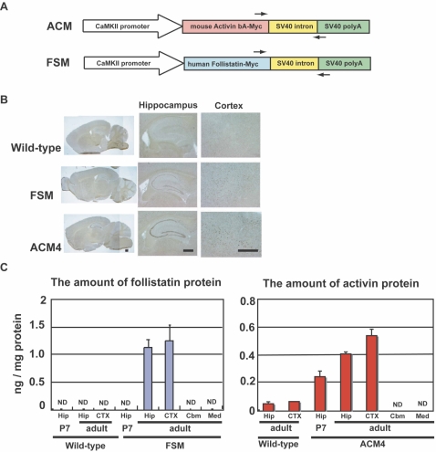
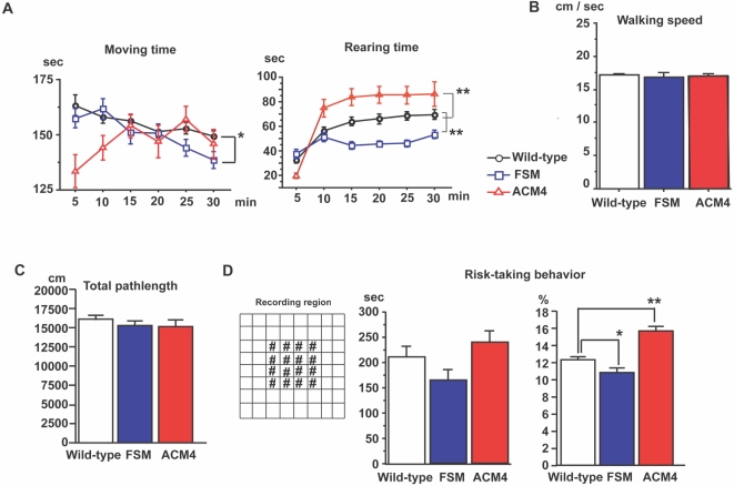
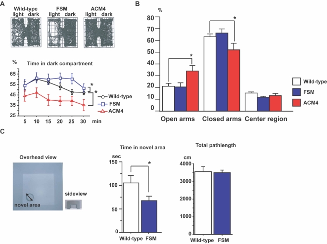
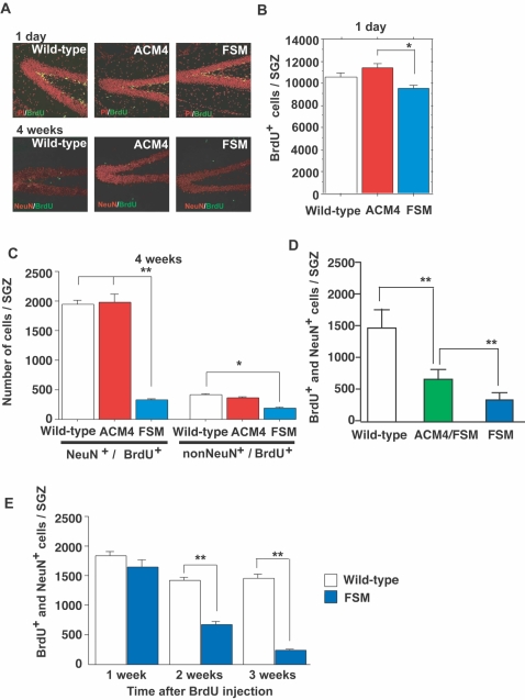
Activin, a member of the transforming growth factor-beta superfamily, is an endocrine hormone that regulates differentiation and proliferation of a wide variety of cells. In the brain, activin protects neurons from ischemic damage. In this study, we demonstrate that activin modulates anxiety-related behavior by analyzing ACM4 and FSM transgenic mice in which activin and follistatin (which antagonizes the activin signal), respectively, were overexpressed in a forebrain-specific manner under the control of the alphaCaMKII promoter. Behavioral analyses revealed that FSM mice exhibited enhanced anxiety compared to wild-type littermates, while ACM4 mice showed reduced anxiety. Importantly, survival of newly formed neurons in the subgranular zone of adult hippocampus was significantly decreased in FSM mice, which was partially rescued in ACM4/FSM double transgenic mice. Our findings demonstrate that the level of activin in the adult brain bi-directionally influences anxiety-related behavior. These results further suggest that decreases in postnatal neurogenesis caused by activin inhibition affect an anxiety-related behavior in adulthood. Activin and its signaling pathway may represent novel therapeutic targets for anxiety disorder as well as ischemic brain injury.


Conflict of interest statement

Competing Interests: The authors have declared that no competing interests exist.

Figures

Figure 1
Figure 1. Generation of transgenic mice and expression analysis of the transgene.
(A) Schematic representation of transgene constructs. Narrow arrows indicate the location and direction of RT-PCR primers (Figure S2). (B) Photographs of typical in situ hybridization are shown. DIG-labeled cRNA probe corresponding to the SV40 polyA sequence was hybridized to sagittal sections of the brain from 16–22 week-old wild-type, FSM and ACM4 mice. Scale bars, 500 µm. (C) Follistatin and activin protein levels in the hippocampus (Hip), cortex (CTX), cerebellum (Cbm) and medulla (Med) in wild-type, FSM and ACM4 mice measured by ELISA. Results are shown as mean±s. e. m. (n = 4) Except for activin in CTX in wild-type (n = 1). Adult indicates 16–22 weeks-old. P7, postnatal day 7.
Figure 2
Figure 2. Activin protein levels in the brain influence locomotor activity.
(A) Statistical analyses of the open field test showing time spent in locomotion and rearing [wild-type littermates (black circles), n = 34; FSM (blue squares), n = 18; ACM4 (red triangles), n = 11]. Each plot represents an average of 5 minutes. *, p<0.05; **, p<0.001; Fisher's test. (B and C) Statistical analyses of walking velocity (B) and total pathlength (C) during 30 minutes of open field test. (D) Risk taking behavior test. Left panel, overhead view of the box used for open field test. #, area defined as a center region. Mid panel, time spent in the center region during the 30 minutes of open field testing. Right panel, the percentage pathlength in the center region. Results are shown as mean±s. e. m.
Figure 3
Figure 3. Activin protein levels in the brain modulate anxiety-related behavior.
(A) Upper panels, typical traces in the light and dark test for each genotype. Lower panels, time (%) spent in the dark compartment was measured over 30 min. Wild-type littermates (black circles), n = 34; FSM (blue squares), n = 18; ACM4 (red triangles), n = 11. *, p<0.05, Fisher's test. (B) Statistical analyses of elevated plus-maze. Wild-type littermates, n = 26; FSM, n = 7; ACM4, n = 11. p values indicate ANOVA for genotype effect. *, p<0.05; t-test. (C) Left panels shows the apparatus used for the novel-area accessing test. Arrows indicate points of entry to the cylinder. Small picture shows side view of the cylinder. Bar graphs show time spent in novel area (inside cylinder) and distance traveled during 10 min testing. *, p<0.05, t-test. Results are shown as mean±s. e. m.
Figure 4
Figure 4. Activin signal is essential for survival of newly generated neurons.
(A) BrdU positive cells (green) in the SGZ. Mice were sacrificed 1 day (upper panels) or 4 weeks (lower panels) after BrdU administration. At 4 weeks, most BrdU-positive cells were co-labeled with NeuN (red), a marker for mature neurons. Propidium iodide (PI, red) was used as a nuclear marker. (B) Number of BrdU-positive cells in the SGZ. The Y-axis indicates the number of BrdU-positive cells of the entire hippocampus. ACM4 had more BrdU-positive cells than FSM (wild-type, n = 21 animals; ACM4, n = 6 animals; FSM, n = 10 animals). (C) Number of BrdU-positive cells differentiated to neurons (NeuN+/BrdU+) or differentiated to another cell type (non-NeuN+/BrdU+) in the SGZ (wild-type, n = 25; ACM4, n = 14; FSM, n = 9). Animals were sacrificed 4 weeks after BrdU administration. (D) Number of BrdU- and NeuN-double positive cells in the SGZ (wild-type, n = 32; ACM4/FSM, n = 9; FSM, n = 14). Animals were sacrificed 4 weeks after BrdU administration. (E) Number of BrdU/NeuN-double positive cells in the SGZ. Animals were sacrificed 1, 2, or 3 weeks after BrdU administration. At the 2-week stage (wild-type, n = 7; FSM, n = 8), but not at the 1-week stage (wild-type, n = 7; FSM, n = 9), the number of double positive cells was significantly decreased in FSM mice compared with wild-type littermates. Error bars indicate the s. e. m. *, p<0.05; **, p<0.01; t-test.

Similar articles

Cited by

References

    1. Cryan JF, Holmes A. The ascent of mouse: advances in modelling human depression and anxiety. Nat Rev Drug Discov. 2005;4:775–790. - PubMed
    1. Kalueff AV, Wheaton M, Murphy DL. What's wrong with my mouse model? Advances and strategies in animal modeling of anxiety and depression. Behav Brain Res. 2007;179:1–18. - PubMed
    1. Wong ML, Licinio J. From monoamines to genomic targets: a paradigm shift for drug discovery in depression. Nat Rev Drug Discov. 2004;3:136–151. - PubMed
    1. Dranovsky A, Hen R. Hippocampal neurogenesis: regulation by stress and antidepressants. Biol Psychiatry. 2006;59:1136–1143. - PMC - PubMed
    1. Earnheart JC, Schweizer C, Crestani F, Iwasato T, Itohara S, et al. GABAergic control of adult hippocampal neurogenesis in relation to behavior indicative of trait anxiety and depression states. J Neurosci. 2007;27:3845–3854. - PMC - PubMed

Publication types

Substances

LinkOut - more resources