

Efficient in vivo electroporation of the postnatal rodent forebrain
- PMID: 18382666
- PMCID: PMC2270900
- DOI: 10.1371/journal.pone.0001883
Efficient in vivo electroporation of the postnatal rodent forebrain
Abstract
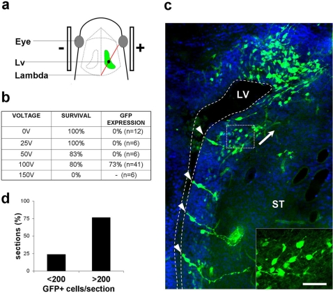
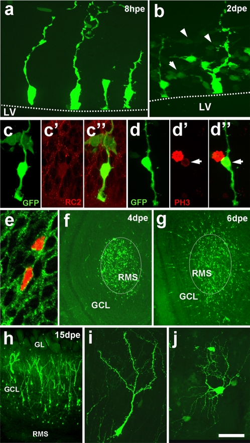
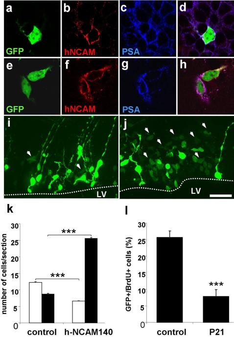
Functional gene analysis in vivo represents still a major challenge in biomedical research. Here we present a new method for the efficient introduction of nucleic acids into the postnatal mouse forebrain. We show that intraventricular injection of DNA followed by electroporation induces strong expression of transgenes in radial glia, neuronal precursors and neurons of the olfactory system. We present two proof-of-principle experiments to validate our approach. First, we show that expression of a human isoform of the neural cell adhesion molecule (hNCAM-140) in radial glia cells induces their differentiation into cells showing a neural precursor phenotype. Second, we demonstrate that p21 acts as a cell cycle inhibitor for postnatal neural stem cells. This approach will represent an important tool for future studies of postnatal neurogenesis and of neural development in general.
Conflict of interest statement
Figures
Similar articles
-
Targeted electroporation of defined lateral ventricular walls: a novel and rapid method to study fate specification during postnatal forebrain neurogenesis.Neural Dev. 2011 Apr 5;6:13. doi: 10.1186/1749-8104-6-13. Neural Dev. 2011. PMID: 21466691 Free PMC article.
-
FoxJ1-expressing cells contribute to neurogenesis in forebrain of adult rats: evidence from in vivo electroporation combined with piggyBac transposon.Exp Cell Res. 2013 Nov 1;319(18):2790-800. doi: 10.1016/j.yexcr.2013.08.028. Epub 2013 Sep 25. Exp Cell Res. 2013. PMID: 24075965
-
Simultaneous expression of different transgenes in neurons and glia by combining in utero electroporation with the Tol2 transposon-mediated gene transfer system.Genes Cells. 2010 May;15(5):501-12. doi: 10.1111/j.1365-2443.2010.01397.x. Epub 2010 Apr 7. Genes Cells. 2010. PMID: 20384787 Free PMC article.
-
Radial glial origin of the adult neural stem cells in the subventricular zone.Prog Neurobiol. 2007 Sep;83(1):24-36. doi: 10.1016/j.pneurobio.2006.11.002. Epub 2006 Dec 29. Prog Neurobiol. 2007. PMID: 17196731 Review.
-
Development and aging of a brain neural stem cell niche.Exp Gerontol. 2017 Aug;94:9-13. doi: 10.1016/j.exger.2016.11.007. Epub 2016 Nov 17. Exp Gerontol. 2017. PMID: 27867091 Free PMC article. Review.
Cited by
-
miR-7a regulation of Pax6 controls spatial origin of forebrain dopaminergic neurons.Nat Neurosci. 2012 Jun 24;15(8):1120-6. doi: 10.1038/nn.3142. Nat Neurosci. 2012. PMID: 22729175
-
Single neuron electroporation in manipulating and measuring the central nervous system.Int Arch Med. 2010 Nov 5;3:28. doi: 10.1186/1755-7682-3-28. Int Arch Med. 2010. PMID: 21054865 Free PMC article.
-
Recent Advances in In Vivo Somatic Cell Gene Modification in Newborn Pups.Int J Mol Sci. 2023 Oct 18;24(20):15301. doi: 10.3390/ijms242015301. Int J Mol Sci. 2023. PMID: 37894981 Free PMC article. Review.
-
Long Non-coding RNA T-UCstem1 Controls Progenitor Proliferation and Neurogenesis in the Postnatal Mouse Olfactory Bulb through Interaction with miR-9.Stem Cell Reports. 2020 Oct 13;15(4):836-844. doi: 10.1016/j.stemcr.2020.08.009. Epub 2020 Sep 24. Stem Cell Reports. 2020. PMID: 32976763 Free PMC article.
-
Harnessing DNA-induced immune responses for improving cancer vaccines.Hum Vaccin Immunother. 2012 Nov 1;8(11):1682-93. doi: 10.4161/hv.22345. Epub 2012 Oct 30. Hum Vaccin Immunother. 2012. PMID: 23111166 Free PMC article. Review.
References
-
- Nakamura H, Katahira T, Sato T, Watanabe Y, Funahashi J. Gain- and loss-of-function in chick embryos by electroporation. Mech Dev. 2004;121:1137–1143. - PubMed
-
- Saito T, Nakatsuji N. Efficient gene transfer into the embryonic mouse brain using in vivo electroporation. Dev Biol. 2001;240:237–246. - PubMed
-
- Mir LM, Moller PH, Andre F, Gehl J. Electric pulse-mediated gene delivery to various animal tissues. Adv Genet. 2005;54:83–114. - PubMed
Publication types
MeSH terms
Substances
LinkOut - more resources
Full Text Sources
Other Literature Sources
